Search This Blog

Friday 4 November 2016

The HDL correlations in CANHEART probably don't mean what the druglords will want them to mean


The CANHEART study findings on HDL have made a big splash, supposedly debunking the idea that raising HDL is a good idea. Of course, raising HDL with drugs by sticking a spanner in the works at some point has never been an effective strategy, and there are genetic polymorphisms that give elevated HDL of little worth, but healthy diet and lifestyle changes that are reasonably expected to extend life always raise HDL a bit. Is this meaningless?
In CANHEART very high HDL cholesterol was actually associated with higher non-cardiovascular mortality.[1]
Especially levels over 90 mg/dl (2.33 mmol/l), but also over 70 mg.dl in men (1.81 mmol/l).
These are very high HDL levels, I don't remember seeing levels this high in non-drinkers on LCHF diets, no matter how much coconut oil they eat.

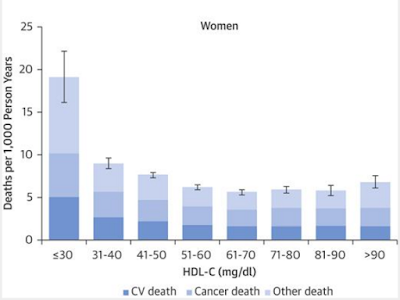


The most obvious question is, what about alcohol? Alcohol elevates HDL but at high intakes promotes secretion of useless and atherogenic HDL subtypes. Ko at al claimed to have adjusted for excess alcohol intake, which was highest in those with highest HDL;

"Heavy alcohol consumption, as defined by the use of 5 or more drinks on 12 or more occasions per year was also included in the model for non-cardiovascular non-cancer death."

Newsflash - drinking 6 drinks 13 times per year will not raise your HDL. You really need to be a chronic alcoholic. In 2012, approximately 5 million Canadians (or 18 % of the population) aged 15 years and older met the criteria for alcohol abuse or dependence at some point in their lifetime, but how many at any one time qualify as chronically alcoholic is unknown.


Even so, this adjustment was far from perfect.

"Since the use of smoking and alcohol was not available in entire CANHEART cohort, we imputed smoking status and heavy alcohol use for those with missing data based on the characteristics of the respondents to the Canadian Community Health Survey. Multiple imputation using complete observations and 10 imputation datasets was conducted. Smoking status was available for 5,093 individuals and alcohol use was available for 5,077 individuals who completed the survey."

This was a tiny fraction of the 631,762 individuals in the study - less than 1% - and presumably was either restricted to a single geographical area, or a few especially obliging subjects.
Alcohol intake is known to be misreported in dietary surveys by a factor of 2-3. Alcoholism is probably under-reported to health professionals to a much greater extent, especially in countries where health insurance is a major factor in access to care.

Another confounder is the effect of genetic hyperalphalipoproteinemia. One genetic cause of very high HDL is a CETP defect.

"...the in vitro evidence showed large CE-rich HDL particles in CETP deficiency are defective in cholesterol efflux. Similarly, scavenger receptor BI (SR-BI) knockout mice show a marked increase in HDL-cholesterol but accelerated atherosclerosis in atherosclerosis-susceptible mice. Recent epidemiological studies in Japanese-Americans and in Omagari area where HALP subjects with the intron 14 splicing defect of CETP gene are markedly frequent, have demonstrated an increased incidence of coronary atherosclerosis in CETP-deficient patients. Thus, CETP deficiency is a state of impaired reverse cholesterol transport which may possibly lead to the development of atherosclerosis."[2]


Ko et al do not mention the likelihood of such conditions affecting their analysis. Even if we assume that both chronic alcoholism and hyperalphalipoproteinemia are rare conditions, men with HDL over 90mg/l were less than 0.3% of the study population, and of these few men, only a few dozen died during the study. The exact number isn't clear because the only mortality data given is for adjusted age-standardized rates per 1,000, but from total deaths and these rates I estimate it to be (at the very most) 70-80 deaths, of which 30-35 were non-cardiovascular and non-cancer deaths, out of about 2240 men. The majority of alcohol-related such deaths in Canada are due to alcoholic liver disease, motor vehicle accidents and alcohol-related suicides. Had Ko et al given a breakdown of non-cardiovascular causes of death for the highest HDL categories, it would have been relatively easy to tell how many of these were due to alcoholism.

Overall, people in the high HDL categories exercised more, had lower triglycerides, less diabetes, lower LDL, more ideal BMI, and ate more fruit and vege than people in the middle and lower ranges.
Did these things cause them to die at a higher rate?
Here's an alternative explanation - the baseline characteristics represent only the vast majority of people in each category.  The vast majority of people in each HDL category, even the highest, didn't die. The people who died in the high HDL categories tended to be the people with alcoholism and poorly-managed genetic hyperalphalipoproteinemia, and their baseline characteristics, had they been isolated, would have been quite different. These are the people for whom high HDL is not protective, and, as their numbers increased in categories of increasing HDL, the usual dose-response relationship between HDL and cardiovascular disease and cancer, seen in better-controlled populations, was lost.


A criticism is that Ko et al have misrepresented the lipid lowering trial data to support their thesis.
They say "Several contemporary studies have shown a lack of significant association of HDL-C levels and outcomes for patients on higher-intensity statins, with coronary artery disease, or who had undergone coronary artery bypass graft surgery (12,13,15)."
However, reference 12 states

"In 8901 (50%) patients given placebo (who had a median on-treatment LDL-cholesterol concentration of 2.80 mmol/L [IQR 2.43-3.24]), HDL-cholesterol concentrations were inversely related to vascular risk both at baseline (top quartile vs bottom quartile hazard ratio [HR] 0.54, 95% CI 0.35-0.83, p=0.0039) and on-treatment (0.55, 0.35-0.87, p=0.0047). By contrast, among the 8900 (50%) patients given rosuvastatin 20 mg (who had a median on-treatment LDL-cholesterol concentration of 1.42 mmol/L [IQR 1.14-1.86]), no significant relationships were noted between quartiles of HDL-cholesterol concentration and vascular risk either at baseline (1.12, 0.62-2.03, p=0.82) or on-treatment (1.03, 0.57-1.87, p=0.97). Our analyses for apolipoprotein A1 showed an equivalent strong relation to frequency of primary outcomes in the placebo group but little association in the rosuvastatin group."[3]

In other words, people in the top quartile for HDL and ApoA1 on placebo had the lowest vascular risk, and these people got no extra benefit from LDL lowering with a statin. And because we are looking at quartiles, not isolating a small number of people who have freakishly high HDL for some reason, there is a true dose-response effect of HDL between quartiles in the placebo arm.
This effect has been seen in multiple trials. Drug trials are likely to exclude alcoholics and binge drinkers.
All these 3 references tell us is that the predictive value of HDL is excellent, but is lost when people are undergoing intensive treatment for coronary artery disease, a classic case of Goodhart's law, "When a measure becomes a target, it ceases to be a good measure." We see this again and again with intensive drug treatment of metabolic markers.
Thankfully, it doesn't seem to apply to diet and lifestyle interventions.


References

[1] Ko DT, Alter DA, Guo H, et al. High-Density Lipoprotein Cholesterol and Cause-Specific Mortality in Individuals Without Previous Cardiovascular Conditions: The CANHEART Study. J Am Coll Cardiol. 2016;68(19):2073-2083. doi:10.1016/j.jacc.2016.08.038.

[2] Yamashita S, Maruyama T, Hirano K, Sakai N, Nakajima N, Matsuzawa Y. 
Molecular mechanisms, lipoprotein abnormalities and atherogenicity of hyperalphalipoproteinemia.
Atherosclerosis. 2000 Oct;152(2):271-85.


[3] Ridker  P.M., Genest  J., Boekholdt  S.M., et al; for the JUPITER Trial Study Group. HDL cholesterol and residual risk of first cardiovascular events after treatment with potent statin therapy: an analysis from the JUPITER trial. Lancet. 2010;376:333-339.
Sunday 25 September 2016

Animal Protein vs Plant Protein - the illusion of scale in diet epidemiology.

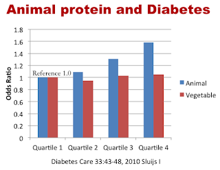
This graph appeared in Jason Fung's excellent Intensive Dietary Management blog here. I don't really want to disagree with Jason's statement that animal protein raises insulin more than plant protein, as I haven't looked into the evidence for that or what it means - I merely want to point out that this graph, and the paper it comes from, do not by themselves provide evidence that eating animal protein is associated with a higher risk of developing type 2 diabetes than eating plant protein.


The paper, by Sluijs et al, is titled "Dietary Intake of Total, Animal, and Vegetable Protein and Risk of Type 2 Diabetes in the European Prospective Investigation into Cancer and Nutrition (EPIC)-NL Study" and states
"During 10 years of follow-up, 918 incident cases of diabetes were documented. Diabetes risk increased with higher total protein (hazard ratio 2.15 [95% CI 1.77–2.60] highest vs. lowest quartile) and animal protein (2.18 [1.80–2.63]) intake. Adjustment for confounders did not materially change these results. Further adjustment for adiposity measures attenuated the associations. Vegetable protein was not related to diabetes. Consuming 5 energy % from total or animal protein at the expense of 5 energy % from carbohydrates or fat increased diabetes risk.
Diets high in animal protein are associated with an increased diabetes risk. Our findings also suggest a similar association for total protein itself instead of only animal sources. Consumption of energy from protein at the expense of energy from either carbohydrates or fat may similarly increase diabetes risk. This finding indicates that accounting for protein content in dietary recommendations for diabetes prevention may be useful."

Leaving aside the implausibility of the finding for the moment, there's an inconsistency in this abstract. If vegetable protein isn't "related to" diabetes, why is total protein a problem?

I knew from reading Song et al recently that the quartiles for vegetable protein actually represent quite small amounts. In the graph above, the upper quartile of vegetable protein is eating 33g/day, while the lower quartile of animal protein is eating 35g/day. So if you want to compare similar amounts of these proteins, you need to compare upper quartile vege with lower quartile animal, and they have exactly the same association with diabetes. And total protein (meat and vege combined) actually had a stronger association with diabetes than animal protein (1.67 in model 3, vs 1.58 for animal protein).

In real life, most people were eating both sorts of protein. Across the animal protein quartiles in Table 1, vegetable protein stayed very constant (people ate much the same amount of wheat). Unfortunately, there is no baseline data that tells us how much animal protein the quartiles of vegetable protein ate.

But let's take a common-sense approach to this data. Model 3, which I cited earlier, isn't adjusted for BMI and waist circumference. The highest protein quartile reports eating fewer total calories than the others, but has significantly greater BMI and waist circumference. And when these are adjusted for (Model 4, Table 2), voila, the association between protein and diabetes disappears from the quartile calculations; only the per 10g association remains. And this, though small, is greater for total protein (1.16) than for animal protein (1.13).
Amount of total protein across quartiles is 64g, 72g, 79g, and 88g. This range hardly seems excessive. Why it would be associated with diabetes at all is, frankly, a mystery. And what would happen if this population ate 64g, 72g, 79g, or 88g of plant protein is completely unknowable.

EDIT: some afterthoughts

The abstract of this paper only reports the completely unadjusted HRs. That is, not even age or sex adjusted. "Attenuated" really means "Disappeared".

The upper protein quartile ate fewer calories, were more active, and had significantly higher BMI (2 points) and waist circumference (4cm).
In other words, either this study breaks the sacred rules of diet thermodynamics, or diet was not reported accurately.
The quartile who ate least vegetable protein had to have eaten more animal protein than the others, just to survive. So why is their risk of diabetes so low?

Monday 19 September 2016

Court of last appeal - the early history of the high-fat diet for diabetes

It's a long story, and not a proud one. Seeing an email in my inbox from the Journal of Diabetes & Metabolism, which seemed like the title of a journal I'd investigated earlier, I impulsively sent off a draft of my history of Louis "Harry" Newburgh and the Michigan diet. I just emailed the unformatted pdf to them, and never engaged any portal or website.
The journal replied, in terrible English, that my article would be accepted with changes requested by one of "two reviewers" supplying a short paragraph each. This request was that I shorten and focus the abstract, and format references.
At this stage I realised this was an OMICs journal, of predatory reputation. I sent off the formatted article with no change but a small edit to the abstract to see what would happen.
The next thing I knew, I was sent an author proof. And an invoice for US$4,000. For some reason the illustration of Newburgh had been titled as being of Frederick Allen, a name which appears in the text but was never attached to any picture. I corrected this and received an authorproof with errors corrected.
Here is this proof, which I consider to be an accurate version of the paper, albeit I believe it was not properly peer-reviewed.

https://www.dropbox.com/s/l3pxp9lazyu5v04/2155-6156-7-696_Authorproof.pdf?dl=0

I never paid the fee, and received numerous reminders, not all addressed to me. I still receive emails from other OMICs journals requesting my input. I was never asked to sign a COI declaration of any sort, nor any other agreement (the work is Open Commons). I had read a thread on researchgate in which an author describes seeing their work published despite not paying the fee.
https://www.researchgate.net/post/Can_I_trust_OMICS_publishing_group

So I searched for my paper online and found it here.
http://www.omicsonline.org/open-access/court-of-last-appeal--the-early-history-of-the-highfat-diet-for-diabetes-2155-6156-1000696.php?aid=78354

Not only is the picture of Newburgh captioned Frederick Lewis Allen 1932, the references in the HTML version (but luckily not the pdf) are imported from some other paper, probably from a different journal.
I feel like this is a punishment to make an example of defaulting authors!

Anyway, the history of Newburgh is published, which is the main thing, but I feel ethically unclean, and whether anyone can ever now cite this article in a proper paper is uncertain. At least OMICs have not claimed exclusive rights in the matter, so republication is not out of the question. But then, if I'd thought this necessarily detailed history would be easy to publish in a proper journal, it would never have ended up in the hands of OMICs.

I'd like to thank Ash Simmonds
 and Zooko for introducing me to the work of Prof Newburgh.

Tuesday 23 August 2016

Evidence of cardiovascular benefits of LCHF diets, despite no change or increase in LDL, from drug trials

A recent meta-analysis of low-carb diets and cardiovascular risk factors found, predictably, that low carb diets decrease triglycerides (TG), increase HDL, and - significantly, on average, but not consistently, and only by a small amount - elevate LDL.
The authors argued that this was not evidence of cardiovascular safety. "Low-carbohydrate diets increase LDL-cholesterol, and thereby indicate increased risk of CVD."
Other cardiologists disputed this (including  Axel F. SigurĂ°sson of the Doc's Opinion blog), citing evidence that TG and HDL are better markers of cardiovascular health than is LDL.[1]
The authors responded with a narrowly focussed argument [2] -

1) Mendelian randomisation shows the genes associated with LDL are associated with CVD, whereas genes associated with HDL are not, and those with TG only slightly.


I think this is faulty logic. Genes are the things we cannot change, so the association of TG and HDL with CVD risk, seen in the baseline characteristics of participants in drug trials (those with high HDL and low TG have low CVD risk in placebo arm and get no extra benefit in LDL-lowering arm  - links to those studies in this post), is probably due to diet and lifestyle factors, as Mendelian randomisation seems to rule out a strong genetic influence; but it does suggest that these factors are downstream markers of some other, more proximal "root cause" factor.


2) Drugs that elevate HDL have no effect on CVD risk, whereas statins, which lower LDL, do have some effect.


As with their point 1), these authors simply did not look deeply enough into the literature. There are many drugs that have lowered LDL with no or harmful effects on CVD outcomes, which seem to have been ignored in this argument. As for HDL, alcohol, for example, is a drug that elevates HDL and decreases CVD risk, see e.g.[3]

However, this link is observational. Better data comes from the trials of a new class of drugs, the SGLT2 inhibitors. Empagliflozin elevates both HDL and LDL. "in T2DM patients with high CVD risk empagliflozin compared to placebo reduced the primary major adverse cardiac event end point (CV death, nonfatal myocardial infarction, nonfatal stroke) by 14%. This beneficial effect was driven by a 38% reduction in CV mortality with no significant decrease in nonfatal myocardial infarction or stroke. Empagliflozin also caused a 35% reduction in hospitalization for heart failure without affecting hospitalization for unstable angina."[4]
Empagliflozin was also shown to be renoprotective, significantly reducing the incidence of worsening nephropathy, by 39%. This is interesting because nephropathy is a vascular pathology of diabetes.

SGLT2 inhibitors mimic the effect of low-carbohydrate ketogenic diets over a wide range of metabolic parameters (increased sodium excretion, decreased extracellular volume, increased HDL and LDL, reduced requirement for insulin, increased ketogenesis). The doctors are still arguing about the mechanism of benefit.

However, we note that 48% of the subjects were receiving insulin at baseline (median daily dose 54 units) and 43% were using sulfonylureas (which increase insulin secretion). During the EMPA-REG trial the rate of addition of new medications was (drug vs placebo) 5.8% vs. 11.5% for insulin and 3.8% vs. 7.0% for sulfonylureas, consistent with studies in which SGLT2 inhibitors decrease insulin requirements in type 1 diabetes.[5]

Are there other drug trials that support this model? The STOP-NIDDM study tested acarbose for the prevention of diabetes in a group of patients with impaired glucose tolerance. Acarbose inhibits the digestion of starch, and side effects of diarroeah  and flatulence limited compliance (how much simpler it would be to simply resist starch).

"211 (31%) of 682 patients in the acarbose group and 130 (19%) of 686 on placebo discontinued treatment early. 221 (32%) patients randomised to acarbose and 285 (42%) randomised to placebo developed diabetes (relative hazard 0.75 [95% CI 0.63-0.90]; p=0.0015). Furthermore, acarbose significantly increased reversion of impaired glucose tolerance to normal glucose tolerance."

Less carbohydrate entering the bloodstream from the gut = less progression of pre-diabetes to diabetes (and hence less CVD risk). It's not rocket science, unless you work for a pharmaceutical company in some capacity.

Acarbose doesn't alter LDL or HDL, but it does decrease triglycerides (thus improve the TG/HDL ratio) and VLDL. It also reduces the atherogenicity of LDL particles.
"The density gradient lipoprotein separation and disk polyacrylamide gel electrophoresis analyses showed that acarbose reduced the amount of small dense LDL, a more atherogenic and oxidatively susceptible form of LDL. We also found that the fatty acid composition of LDL changed after the treatment: polyunsaturated (omega-3) fatty acid, a beneficial substance for preventing cardiovascular disease, was significantly increased, whereas saturated fatty acids and triglyceride were decreased in the LDL of the acarbose-treated group."[7]
Decrease in sdLDL and serum SFAs is also an effect of low carb diets.

Does acarbose lower CVD incidence? You bet it does. In a meta-analysis of 7 RCTs of acrabose vs placebo in patients with T2DM, "The treatment significantly reduced the risk for ‘myocardial infarction’ (hazards ratio=0.36 [95% Cl 0.16–0.80], P=0.0120) and ‘any cardiovascular event’ (0.65 [95% Cl 0.48–0.88], P=0.0061)."[8]

In an experiment in fructose fed rats, there was no difference in blood glucose, but fructose increased, and acarbose subsequently reduced, insulin levels.[9]
In a double-blind, placebo-controlled, randomised cross-over study in subjects (n=10) with type 1 diabetes, "Acarbose produced a statistically significant reduction in mean insulin requirement over a 3-hr period following the meal compared with placebo (5171.7+/-2282.6 mU vs 8074.5+/-3045.4 mU; p=0.003). The level of blood glucose control over the same period was similar in the two groups.".


We measure fasting glucose, HbA1c, and OGTT glucose response to diagnose type 2 diabetes because these are easy and cheap to measure, but if we could measure the insulin response as easily and cheaply we would have a better guide to risk of complications and CVD and to the type and stage of diabetes.

This is because most of the pathologies of type 2 diabetes - cardiovascular disease and vascular disease in particular, but also, probably, the progression of beta-cell failure - are driven by elevated insulin levels.[11]
On the other hand, drugs that reduce both glucose and insulin (secretion or requirement) by restricting uptake or increasing excretion of glucose - i.e. acarbose or SGLT2 inhibitors (EMPA-REG trial) - significantly reduce the risk of cardiovascular disease and vascular pathologies.
What of statins? These have some lesser effect on the incidence of cardiovascular and vascular disease, despite the potential for increased blood glucose.
Statins inhibit the synthesis of cholesterol in cells, and the synthesis of excessive cholesterol, which disrupts mitochondrial function, is driven by excessive insulin concentrations.
"β-Hydroxy-β-methylglutaryl coenzyme A reductase activity in rat liver increased 2 to 7-fold after subcutaneous administration of insulin into normal or diabetic animals. Reductase activity began increasing after one hour, rose to a maximum in two to three hours, and then declined to the control level after six hours. This response was elicited during the time of day when the normal diurnal variation in reductase activity approached a minimum. It was also elicited when animals did not have access to food. This stimulation of reductase activity was completely blocked when glucagon was administered in conjunction with insulin. The increase in reductase activity after insulin administration was accompanied by a proportionate increase in activity for the conversion of acetate to cholesterol."[12]
What therapy lowers the secretion of or requirement for insulin, but does not increase and will usually lower blood glucose?
A low carbohydrate, high fat diet.
Q.E.D.

[1] Thomas R. Wood, Robert Hansen, Axel F. SigurĂ°sson and GuĂ°mundur F. JĂ³hannsson (2016). The cardiovascular risk reduction benefits of a low-carbohydrate diet outweigh the potential increase in LDL-cholesterol. British Journal of Nutrition, 115, pp 1126-1128. doi:10.1017/S0007114515005450.


[2] Nadia Mansoor, Kathrine J. Vinknes, Marit B. Veierød and Kjetil Retterstøl (2016). Low-carbohydrate diets increase LDL-cholesterol, and thereby indicate increased risk of CVD. British Journal of Nutrition, 115, pp 2264-2266. doi:10.1017/S0007114516001343.


[3] Roles of Drinking Pattern and Type of Alcohol Consumed in Coronary Heart Disease in Men

Kenneth J. Mukamal, M.D., M.P.H., Katherine M. Conigrave, M.B., B.S., Ph.D., Murray A. Mittleman, M.D., Dr.P.H., Carlos A. Camargo, Jr., M.D., Dr.P.H., Meir J. Stampfer, M.D., Dr.P.H., Walter C. Willett, M.D., Dr.P.H., and Eric B. Rimm, Sc.D.
N Engl J Med 2003; 348:109-118January 9, 2003DOI: 10.1056/NEJMoa022095


[4] SGLT2 Inhibitors and Cardiovascular Risk: Lessons Learned From the EMPA-REG OUTCOME Study.

Muhammad Abdul-Ghani, Stefano Del Prato, Robert Chilton and Ralph A. DeFronzo.
Diabetes Care 2016 May; 39(5): 717-725.

[5] https://www.wikijournalclub.org/wiki/EMPA-REG_OUTCOME


[6] Lancet. 2002 Jun 15;359(9323):2072-7.

Acarbose for prevention of type 2 diabetes mellitus: the STOP-NIDDM randomised trial.
Chiasson JL1, Josse RG, Gomis R, Hanefeld M, Karasik A, Laakso M; STOP-NIDDM Trail Research Group.

[7]  Acarbose ameliorates atherogenecity of low-density lipoprotein in patients with impaired glucose tolerance.

Inoue I, Shinoda Y, Nakano T, Sassa M, Goto S, Awata T, Komoda T, Katayama S.
Metabolism. 2006 Jul;55(7):946-52.

[8] Drugs Exp Clin Res. 2005;31(4):155-9.

Acarbose, an alpha-glucosidase inhibitor, improves insulin resistance in fructose-fed rats.
Nakamura K, Yamagishi S, Matsui T, Inoue H.

[9] Diabetes Nutr Metab. 2000 Feb;13(1):7-12.

Influence of acarbose on post-prandial insulin requirements in patients with Type 1 diabetes.
Juntti-Berggren L, Pigon J, Hellström P, Holst JJ, Efendic S.

[10] Acarbose reduces the risk for myocardial infarction in type 2 diabetic patients: meta-analysis of seven long-term studies

M. Hanefeld, M. Cagatay, T. Petrowitsch, D. Neuser, D. Petzinna, M. Rupp.
European Heart Journal. Volume 25, Issue 1. Pp. 10 - 16

[11] Exposure to excess insulin (glargine) induces type 2 diabetes mellitus in mice fed on a chow diet.

Xuefeng Yang, Shuang Mei, Haihua Gu, Huailan Guo, Longying Zha, Junwei Cai, Xuefeng Li, Zhenqi Liu and Wenhong Cao.
Journal of Endocrinology (2014) 221, 469–480

[12] Stimulation by insulin of rat liver β-hydroxy-β-methylglutaryl coenzyme A reductase and cholesterol-synthesizing activities.

M.R. Lakshmanan, Carl M. Nepokroeff, Gene C. Ness, Richard E. Dugan, John W. Porter. Biochemical and Biophysical Research Communications. Volume 50, Issue 3, 5 February 1973, Pages 704-710


Thursday 18 August 2016

Glucokinase mutations, diabetic complications, and cardiovascular disease

This is a very interesting study that was posted by Richard Lehman on his BMJ blog a few years ago. It contains much food for thought.
People with this mis-sense mutation in the gene that encodes glucokinase (GCK), part of the pancreatic beta cell glucose sensor, basically have their sugar thermostat, their glucostat, set too high. They don't produce insulin in response to blood glucose in the pre-diabetic range. In this study, average HbA1c is 6.9%. But the incidence of insulin resistance, obesity, dyslipdaemia, and hypertension in this population is the same as in the normal controls, who have average HbA1c of 5.8% here.
So basically we are looking at mild hyperglycaemia without hyperinsulinaemia and its sequelae.
I think this is a good model for people with type 2 diabetes who have reversed the disease to a pre-diabetic level on a low carb diet, lost weight, and corrected hypertension. No carbs = low insulin, so how much of a problem is mild hyperglycaemia if it persists?
Also, do some people diagnosed with T2DM or prediabetes who go low carb have the GCK mutation without knowing it, meaning they will not get normal blood sugars?


JAMA. 2014 Jan 15;311(3):279-86. doi: 10.1001/jama.2013.283980.
Prevalence of vascular complications among patients with glucokinase mutations and prolonged, mild hyperglycemia.
Steele AM, Shields BM, Wensley KJ, Colclough K, Ellard S, Hattersley AT.

IMPORTANCE:
Glycemic targets in diabetes have been developed to minimize complication risk. Patients with heterozygous, inactivating glucokinase (GCK) mutations have mild fasting hyperglycemia from birth, resulting in an elevated glycated hemoglobin (HbA1c) level that mimics recommended levels for type 1 and type 2 diabetes.

OBJECTIVE:
To assess the association between chronic, mild hyperglycemia and complication prevalence and severity in patients with GCK mutations.

DESIGN, SETTING, AND PARTICIPANTS:
Cross-sectional study in the United Kingdom between August 2008 and December 2010. Assessment of microvascular and macrovascular complications in participants 35 years or older was conducted in 99 GCK mutation carriers (median age, 48.6 years), 91 nondiabetic, familial, nonmutation carriers (control) (median age, 52.2 years), and 83 individuals with young-onset type 2 diabetes (YT2D), diagnosed at age 45 years or younger (median age, 54.7 years).

MAIN OUTCOMES AND MEASURES:
Prevalence and severity of nephropathy, retinopathy, peripheral neuropathy, peripheral vascular disease, and cardiovascular disease.

RESULTS:
Median HbA1c was 6.9% in patients with the GCK mutation, 5.8% in controls, and 7.8% in patients with YT2D. Patients with GCK had a low prevalence of clinically significant microvascular complications (1% [95% CI, 0%-5%]) that was not significantly different from controls (2% [95% CI, 0.3%-8%], P=.52) and lower than in patients with YT2D (36% [95% CI, 25%-47%], P<.001). Thirty percent of patients with GCK had retinopathy (95% CI, 21%-41%) compared with 14% of controls (95% CI, 7%-23%, P=.007) and 63% of patients with YT2D (95% CI, 51%-73%, P<.001). Neither patients with GCK nor controls required laser therapy for retinopathy compared with 28% (95% CI, 18%-39%) of patients with YT2D (P<.001). Neither patients with GCK patients nor controls had proteinuria and microalbuminuria was rare (GCK, 1% [95% CI, 0.2%-6%]; controls, 2% [95% CI, 0.2%-8%]), whereas 10% (95% CI, 4%-19%) of YT2D patients had proteinuria (P<.001 vs GCK) and 21% (95% CI, 13%-32%) had microalbuminuria (P<.001). Neuropathy was rare in patients with GCK (2% [95% CI, 0.3%-8%]) and controls (95% CI, 0% [0%-4%]) but present in 29% (95% CI, 20%-50%) of YT2D patients (P<.001). Patients with GCK had a low prevalence of clinically significant macrovascular complications (4% [95% CI, 1%-10%]) that was not significantly different from controls (11% [95% CI, 5%-19%]; P=.09), and lower in prevalence than patients with YT2D (30% [95% CI, 21%-41%], P<.001).

CONCLUSIONS AND RELEVANCE:

Left columns - GCK, Middle columns - normal, Right columns T2D
Complications, left to right, microvascular, retinopathy, macrovascular.
Despite a median duration of 48.6 years of hyperglycemia, patients with a GCK mutation had low prevalence of microvascular and macrovascular complications. These findings may provide insights into the risks associated with isolated, mild hyperglycemia.


BAM! as they say. Without high insulin, glucose at this level doesn't damage the blood vessels any more than "normal" BG does in a population with "normal" insulin responses to carbohydrate.
It does damage the eyes (but not the nerves), probably because the polyol pathway is insulin-independent, but the rate of retinopathy is already high, at 14%, in the "normal" population. Neuropathy has both a glycotoxic and a microvascular pathology, so is more dependent on hyperinsulinaemia than retinopathy.

A feature of GCK mutation is that blood glucose is highest in the most overweight individuals; this seems to show increased FFA flux boosting gluconeogenesis, or some extra effect of NAFLD increasing insulin resistance.

This is from a paper comparing a sample with the GCK mutation with their normal, non-diabetic family members.[1]
"In subjects with the mutation, beta cell function was impaired, being geometric mean 63 % (normal-100 %) compared with 126 % in the subjects without the mutation (p less than 0.001) measured by HOMA and in a subset assessed by CIGMA 59 % and 127 % (p less than 0.01 ), respectively. There was no difference in fasting insulin concentrations, insulin sensitivity, lipid concentrations or blood pressure between the groups. The haemoglobin A was raised (mean 6.5 % compared with 5.5 % in the subjects without the mutation), but microvascular and macrovascular complications were uncommon."

The authors of the first paper think this is a model for glycaemic control that attains recommended HbA1c targets for T1D and T2D. I don't think this can be the case if extra insulin or sulfonylureas are being used to meet these targets because the diet is still high in carbs. It is a model for the early stages of dietary control of diabetes, with reduced insulin levels or requirements and HbA1c trending down, and weight and blood pressure normalising.

The mechanisms that cause vascular disease in diabetes, including smooth muscle cell dysfunction and impaired eNOS signalling, are the same ones that are supposed to initiate atherosclerosis, whatever the role of lipoproteins in its development. Say it again - it's the insulin stupid.

[1] Diabet Med. 1995 Mar;12(3):209-17.
Clinical characteristics of subjects with a missense mutation in glucokinase.
Page RC1, Hattersley AT, Levy JC, Barrow B, Patel P, Lo D, Wainscoat JS, Permutt MA, Bell GI, Turner RC.


Sunday 7 August 2016

Problems with Song et al Animal Protein vs Plant Protein study

According to Harvard, this truck has saved more lives than an ambulance.

Here we have another study from the hydra-headed monster that is the Harvard school of public health's interpretation of the NHS and HPFS studies. By my count there have been four of these so far this year, all saying much the same thing, that dietary guidelines were correct. Or rather, they've been presented as saying that, even though the last paper, on fat and mortality, found that higher fat intake was associated with reduced mortality. Harvard didn't report that finding in their press release.

There are a number of methodological flaws in all these studies, and they are worth highlighting.
Firstly, the authors have combined two somewhat heterogenous cohort studies, previously published separately, and which present different findings, into what they now call one cohort.
Another way of describing this method is to say that they have cherry-picked two studies to put together. There are other studies that they could have combined with HPfS, or with NHS, to dilute or amplify their results. Of course they chose these studies because they are in charge of both of them, but nonetheless this is probably a unique proceeding.

Secondly, the results are now presented as person-years. This creates a larger number which looks impressive, but obscures the actual n= in each result.

Thirdly, the validity of the data is more questionable than the authors admit. Respondents were asked to estimate how many times they had eaten listed foods on average in the past year. The only verification seems to have been a comparison between a sample of the respondents completing both the FFQ and a 7-day food diary.

"In each FFQ, participants were asked how often, on average, they consumed a standardized portion size of each food during the previous year."
"The Spearman correlation coefficient of intake assessed by the FFQs and 7-day dietary record was 0.56 for animal protein and 0.66 for plant protein."

A Spearman correlation of 1 would have meant that the results were identical. 0.56 may be considered "high validity" in diet epidemiology, but wouldn't be accepted at the vehicle testing station. The results are meant to estimate 365 days not 7 days, so this comparison was incomplete.
So incomplete that the NHS cohort (the female half of this population) has reported eating 1,500 kcal/day on average for many years by the FFQ system.

"Among participants who returned baseline questionnaires, we excluded those who had a history of cancer (except nonmelanoma skin cancer), CVD, or diabetes at baseline, left more than 10 items blank on the baseline FFQ in the NHS and more than 70 items blank in the HPFS, or reported implausible energy intake levels (under 500 or over 3500 kcal/d for women, or under 800 or over 4200 kcal/d for men)."

This seems to state that respondents who seriously under- or over-stated energy intake were still included in the two studies.

Those are objections that pertain to the studies as a whole, but what of the specific findings of this study?

"Of the 131 342 participants, 85 013 were women (64.7%) and 46 329 were men (35.3%) (mean [SD] age, 49 [9] years). The median protein intake, as assessed by percentage of energy, was 14% for animal protein (5th-95th percentile, 9%-22%) and 4% for plant protein (5th-95th percentile, 2%-6%). After adjusting for major lifestyle and dietary risk factors, animal protein intake was weakly associated with higher mortality, particularly cardiovascular mortality (HR, 1.08 per 10% energy increment; 95% CI, 1.01-1.16; P for trend = .04), whereas plant protein was associated with lower mortality (HR, 0.90 per 3% energy increment; 95% CI, 0.86-0.95). These associations were confined to participants with at least 1 unhealthy lifestyle factor based on smoking, heavy alcohol intake, overweight or obesity, and physical inactivity, but not evident among those without any of these risk factors. Replacing animal protein of various origins with plant protein was associated with lower mortality. In particular, the HRs for all-cause mortality were 0.66 (95% CI, 0.59-0.75) when 3% of energy from plant protein was substituted for an equivalent amount of protein from processed red meat, 0.88 (95% CI, 0.84-0.92) from unprocessed red meat, and 0.81 (95% CI, 0.75-0.88) from egg.

There are two things that should jump out here. The first is that intakes of animal protein and plant protein differ by a factor of 3. Most people on LCHF and paleo diets are eating more plant protein than the people in NHS and HPFS cohorts. For the people in the lowest quintile of plant protein, this supplied 2.6% of energy. That's consistent with bread and processed meat being the main sources of plant protein. (wheat is 14% protein, most cheap commercial sausages contain wheat and soy protein. I'm not sure if Song et al factored this latter into their analysis).
The comparison between high and low plant protein intake is between median 2.6%E (about 10 grams of protein for NHS) and 6.6%E (about 25 grams). 25 grams is associated with less mortality than 10g. Neither amount is sufficient to sustain life.
In the animal protein stakes, median of lowest quintile is 8.9%E and highest is 20%E, and this is a range of protein intake consistent with life.
We're not really comparing like with like.

The second thing that jumps out is this:
"These associations were confined to participants with at least 1 unhealthy lifestyle factor based on smoking, heavy alcohol intake, overweight or obesity, and physical inactivity, but not evident among those without any of these risk factors."
This screams "residual confounding". If your associations disappear when you minimise confounding variables, you probably haven't measured or adjusted for these properly.
To their credit, Song et al do recognise this;
"First, given the remaining variation of health behaviors across protein intake categories in the unhealthy-lifestyle group, residual confounding from lifestyle factors may contribute to the observed protein-mortality associations. However, our results are robust to adjustment for a wide spectrum of potential confounders and the propensity score. "
This seems to be saying that because they performed adjustments, and this produced consistent results, therefore those results are likely to be correct.
However, in the last paper by this group based on the exact same data sets, there was evidence of residual confounding, in the form of a positive correlation between respiratory disease mortality and saturated fat (HR 1.56; 95% CI, 1.30-1.87). Saturated fat consumption was associated with a higher incidence of smoking, but this had been adjusted for.

This finding was described as "novel", because it had no support in the literature. Respiratory disease mortality is usually associated with smoking (which was controlled for) and other air quality factors (passive smoking and traffic proximity) which were not.

There are two possible explanations for this correlation.

Either saturated fat strongly increases respiratory mortality via unknown mechanisms which are only operative in doctors and nurses living in the USA in 1980-2012, or,

Doctors and nurses living in the USA between 1980 and 2012, years of strong anti-smoking campaigns and adjusted insurance premiums, are more likely to underreport smoking than the other, non-medical populations in other cohort studies.

I leave it to you to judge which of these explanations is more ontologically parsimonious.

[Edit 18/04/17; Wang et al have confirmed that the low SFA quintile was 15 years older than the high SFA quintile, due to their cumulative method and changes made by health-conscious individuals. This introduces a larger possibility of both unmeasured confounding and over-adjustment than suggested by baseline data. Individuals have moved between quintiles since baseline, rendering baseline data useless.]

Let us, for arguments sake, take the results at face value; there is no harm from eating extra animal protein from mixed sources instead of carbohydrate, especially if you don't eat commercial crap, and some benefit from eating plant protein (probably from its richer, higher fat sources, as only these will supply extra protein in replacement for carbohydrate).

Imagine a diet where you replace carbohydrate from wheat flour with protein from almond flour. Why, such a diet will reduce your chances of dying, according to Harvard!


Saturday 9 July 2016

This little piggy had none: are fat soluble micronutrients diluted by serum lipids in CVD and psoriasis?

This will be a rambling post, I'm afraid, and more of a sketch of an idea rather than a pinning down.

This excellent pig study, first tweeted by Prof Andro of the Suppversity blog, is clear proof that the Seven Countries study, as long suspected, was severely confounded by latitude, sunlight exposure, and vitamin D.Summary


4 groups of swine (n=16) fed 2 different atherogenic diets, one with 1500iu vs 500iu vit D3, the other with 1,300iu vs no vit D (calcium was supplemented when level dropped too much). for 12 months.
Diet was (as far as I can tell) high sugar (30-50%), high saturated fat (~40% cocoa butter and ghee), with added cholesterol and cholate, plus 8% and 9% chocolate (sic).
Difference between the 2 diets was vitamin D, and may also have been refined versus semi-refined.
Swine fed diet 1 plus 500iu D3 had marked atherosclerosis, swine fed diet 1 plus 1,500iu had very mild changes. Swine fed diet 2 plus zero vit D3 had severe atherosclerosis, swine fed diet 2 plus 1,300iu D3, well this little piggy had none.

(image borrowed from Fat Emperor blog of Ivor Cummings)
The higher the serum cholesterol, the healthier the arteries. Healthiest swine had cholesterol of 406 +/- 34.8 mg/dL, sickest of 352 +/- 33.8 mg/dL, on same atherogenic diet 2.
As a footnote, the rodent version of this company's atherogenic diet (high sugar, high SFA) has "will not cause obesity" on its webpage. Of course not - it would need to supply more PUFA for that to happen.

Anyway, here we have 1,300-1,500iu of vitamin D3 preventing atherosclerosis in pigs (a reasonably human-compatible model, as anyone who's used porcine insulin will attest) weighing 47-57 Kg, making an equivalent human dose a little higher. To get this much vitamin D3 without supplements you'd need to eat lots of salmon (at least 300g/day) or get some sun.

I get psoriasis in winter, just a touch, a few cm but not nice to be itchy. It fades and heals in the summer. I looked up whether it was related to my high cholesterol and it is, it has its own cholesterol pathology and correlation with CVD (but only significant if it covers a larger area than I get).

Recently I decided to supplement vit D3 again - it's midwinter here. I took 10,000iu week on, week off, to get my levels up. After the first week I noticed my psoriasis had stopped itching and was healing. Now I take 6,000iu/day and it's still good.

I found this study using a whopping 35,000iu/day long term for psoriasis and vitiligo.

Dermatoendocrinol. 2013 Jan 1; 5(1): 222–234.
A pilot study assessing the effect of prolonged administration of high daily doses of vitamin D on the clinical course of vitiligo and psoriasis
Danilo C Finamor, Rita Sinigaglia-Coimbra, Luiz C. M. Neves, Marcia Gutierrez, Jeferson J. Silva, Lucas D. Torres, Fernanda Surano, Domingos J. Neto, Neil F. Novo, Yara Juliano, Antonio C. Lopes, and Cicero Galli Coimbra.

Great stuff - fortune favours the brave, and this worked. To tolerate such a high dose of D3, the participants had to restrict dietary calcium. (avoiding dairy products and calcium-enriched foods like oat, rice or soya “milk”) and drink at least 2.5L of fluid per day. Remember, the vitamin D deficient pigs needed to have calcium supplemented after 6 months.

The PASI score significantly improved in all nine patients with psoriasis. Fourteen of 16 patients with vitiligo had 25–75% repigmentation. Serum urea, creatinine and calcium (total and ionized) did not change and urinary calcium excretion increased within the normal range. High-dose vitamin D3 therapy may be effective and safe for vitiligo and psoriasis patients.

So why were such high doses of D3 needed? Fat soluble vitamins are carried to cells on lipid particles, especially LDL, as noted by Doll and Petit in The Causes of Cancer, 1981.

Maret Traber of the Linus Pauling Institute has recently found that high levels of cholesterol and triglycerides reduce the availability of vitamin E to cells.

" In the continuing debate over how much vitamin E is enough, a new study has found that high levels of blood lipids such as cholesterol and triglycerides can keep this essential micronutrient tied up in the blood stream, and prevent vitamin E from reaching the tissues that need it.


The research, just published in the American Journal of Clinical Nutrition, also suggested that measuring only blood levels may offer a distorted picture of whether or not a person has adequate amounts of this vitamin, and that past methods of estimating tissue levels are flawed.

The findings are significant, the scientists say, because more than 90 percent of the people in the United States who don’t take supplements lack the recommended amount of vitamin E in their diet.

Vitamin E is especially important in some places such as artery walls, the brain, liver, eyes and skin, but is essential in just about every tissue in the body. A powerful, fat-soluble antioxidant, it plays important roles in scavenging free radicals and neurologic function. In the diet, it’s most commonly obtained from cooking oils and some vegetables."

And there you have the big confounder in studies that suggest that PUFAs from vegetable oils reduce the risk of CVD and other diseases (including neurological causes of death in the recent NHS and HPFS update). These oils are major sources of vitamin E, but so are nuts, and nuts are associated with the same protection, except better, in relatively small amounts.

This research raises particular concern about people who are obese or have metabolic syndrome,” said Traber, who is the Helen P. Rumbel Professor for Micronutrient Research in the College of Public Health and Human Sciences at Oregon State University, and a principal investigator in OSU’s Linus Pauling Institute.

“People with elevated lipids in their blood plasma are facing increased inflammation as a result,” Traber said. “Almost every tissue in their body is under oxidative attack, and needs more vitamin E. But the vitamin E needed to protect these tissues is stuck on the freeway, in the circulatory system. It’s going round and round instead of getting to the tissues where it’s needed.”

This research was done with 41 men and women, including both younger and older adults, who obtained vitamin E by eating deuterium-labeled collard greens, so the nutrient could be tracked as it moved through the body. Of some interest, it did not find a significant difference in absorption based solely on age or gender. But there was a marked difference in how long vitamin E stayed in blood serum, based on higher level of lipids in the blood – a more common problem as many people age or gain weight."
From an earlier review of vitamin E metabolism and function, by Brigelius-FlohĂ© and Traber:

Similarly, vitamin E deficiency anemia occurs, largely in premature infants, as a result of free radical damage (47). Diminished erythrocyte life span (48, 49) and increased susceptibility to peroxide-induced hemolysis are apparent not only in severe deficiency, but also in marginal vitamin E deficiency in
hypercholesterolemic subjects (50).
Ref 50 is 
Simon, E., Paul, J. L., Atger, V., Simon, A., and Moatti, N.
(1998) Erythrocyte antioxidant status in asymptomatic hypercholesterolemic men. Atherosclerosis 138, 375–381

I have rambled quite long enough. Look beyond the antioxidant focus of what Maret Traber says; fat soluble vitamins and antioxidants are also modifiers of inflammatory responses, endothelial function, and clotting cascades. We know that lipids are sometimes raised by factors that also cause inflammation independently, and that high cholesterol can often be found together with longevity.

If Traber's findings apply to the other fat-soluble vitamins and antioxidants as well, then we have an explanation for the inconsistencies in the relationship between LDL and the risk of CVD and other diseases. In particular, if LDL is elevated by a diet supplying more of these nutrients, it is likely to be healthier than the elevation of triglycerides and perhaps LDL by a diet that doesn't supply as many; a grain-based, refined sugar, low fat diet. Thus the nutrient density of fatty foods and the vegetables consumed with them becomes important. And so does the sun, and our ability to find vitamin D3 in winter.


Thursday 9 June 2016

Atkins, ketones, methylglyoxal and cancer


What you lose on the swings you make up for on the roundabouts.


Recently this study enjoyed a bit a revival as it was used in a presentation at a DAA meet. I'm not sure of the exact context but the Dietitians Association of Australia has been outstandingly fossilised in its attitude to low carb diets.

Ann N Y Acad Sci. 2005 Jun;1043:201-10.

Ketosis leads to increased methylglyoxal production on the Atkins diet.
Beisswenger BG, Delucia EM, Lapoint N, Sanford RJ, Beisswenger PJ.

Abstract
In the popular and widely used Atkins diet, the body burns fat as its main fuel. This process produces ketosis and hence increased levels of beta-hydroxybutyrate (BOB) acetoacetate (AcAc) and its by-products acetone and acetol. These products are potential precursors of the glycotoxin methylglyoxal. Since methylglyoxal and its byproducts are recognized as a significant cause of blood vessel and tissue damage, we measured methylglyoxal, acetone, and acetol in subjects on the Atkins diet. We found that by 14-28 days, methylghyoxal levels rose 1.67-fold (P = 0.039) and acetol and acetone levels increased 2.7- and 6.12-fold, respectively (P = 0.012 and 0.028). Samples from subjects with ketosis showed even greater increases in methylglyoxal (2.12-fold), as well as acetol and acetone, which increased 4.19- and 7.9-fold, respectively; while no changes were seen in samples from noncompliant, nonketotic subjects. The increase in methylglyoxal implies that potential tissue and vascular damage can occur on the Atkins diet and should be considered when choosing a weight-loss program.

Glycation is the major cause of neurological, optic, tissue and vascular damage in diabetes. Glucose, fructose, and methylglyoxal are precursors of advanced glycation endproducts (AGEs). Glycation of proteins creates free-radical generating hotspots. Amongst other things, almost all bad, this does at least serve the function of keeping further excess substrate out of cells.

"Glycation has the potential to alter the biological structure and function of the serum albumin protein. Once it is glycated, it is less efficient for carrying long chain fatty acid.

In experimental model of adipocyte cell lines, albumin-derived AGE has been shown to trigger the generation of intracellular reactive oxygen species leading to an inhibition of glucose uptake."

http://www.ncbi.nlm.nih.gov/pmc/articles/PMC3951818/
Chris Masterjohn has written at length about methylglyoxal pathways here. Suffice to say that there are pathways to clear methylglyoxal, and that the effects of glycation, shown by elevated HbA1c, neuropathy, and microvascular complications have never so far as I know been reported in persons on ketogenic diets. That is, HbA1c in type 2 diabetics drops sharply on a ketogenic diet, but can rise in non-diabetics, though only within the normal range.


One reason for thinking that ketogenic diets are healthy is the Warburg effect. Otto Warburg won the Nobel Prize in 1931 for "discovery of the nature and mode of action of the respiratory enzyme". In 1924 he postulated the Warburg theory of cancer, which (according to Wikipedia) "postulates that the driver of tumorigenesis is an insufficient cellular respiration caused by insult to mitochondria. The term Warburg effect describes the observation that cancer cells, and many cells grown in-vitro, exhibit glucose fermentation even when enough oxygen is present to properly respire. In other words, instead of fully respiring in the presence of adequate oxygen, cancer cells ferment. The Warburg hypothesis was that the Warburg effect was the root cause of cancer. The current popular opinion is that cancer cells ferment glucose while keeping up the same level of respiration that was present before the process of carcinogenesis, and thus the Warburg effect would be defined as the observation that cancer cells exhibit glycolysis with lactate secretion and mitochondrial respiration even in the presence of oxygen."

The ketogenic diet is proposed, and used, as a cancer therapy because it limits exposure to glucose and fructose, which cancer cells can use via the Warburg (and reverse Warburg) effect, and replaces a large part (up to half) of the glucose requirement with ketone bodies, which most tumours cannot easily use.
But what about methylglyoxal? Can cancer cells use methylglyoxal?

No. Methylglyoxal is cytotoxic, without being much of an energy substrate.
A novel mechanism of methylglyoxal cytotoxicity in prostate cancer cells. Link
 Antognelli C, Mezzasomaa L, Fettucciari K, Talesa VN.
The International Journal of Biochemistry & Cell Biology

Volume 45, Issue 4, April 2013, Pages 836–844The results suggest that this physiological compound merits investigation as a potential chemo-preventive/-therapeutic agent, in differently aggressive prostate cancers.

Here's a summary of methylglyoxal cancer research, which includes a human trial. The trial report is linked here.

Do levels of methylglyoxal on a ketogenic diet equal those used in the trial? Probably not. But a ketogenic diet both removes much of the glycolytic fuel that cancers prefer, and replaces it with ketones which they (mostly) can't use, and which is liable to turn into methylglyoxal, which is deadly poison to them.

A useful way of looking at these things is to compare cancer risk in type 1 and type 2 diabetes. Both are exposed to similar levels of excess glucose, but people with type 1 diabetes are occasionally exposed to higher ketone, and thus methylglyoxal, levels (I'm talking about the usual loose management of these conditions, not people on low carb diets).
"It turns out that the types of cancer that are elevated among type 1 diabetes patients are pretty much the same as those that are elevated among type 2 diabetes patients, and the elevation among type 1 diabetes patients is somewhat smaller than the elevation found among type 2 diabetes patients." Link

A ketogenic diet is great for making people metabolically healthy. I don't see why this would result in greater longevity compared to other people who are metabolically healthy. It's a way of catching up, not necessarily of racing ahead.


Tuesday 7 June 2016

#Context - Butter, eggs, and the epidemiology of cardiovascular disease and diabetes


When Ancel Keys started work on his hypothesis, in 1955, he reported that butter only accounted for 4.8% of fats consumed in the USA.[1] Remember that.

It’s well-known that eggs are associated with type 2 diabetes in the USA, but there’s no such association in the rest of the world, and in Finland eggs have protective association with type 2 diabetes.

“When stratified by geographic area, there was a 39% higher risk of DM (95% CI: 21%, 60%) comparing highest with lowest egg consumption in US studies (I2 = 45.4%, P = 0.089) and no elevated risk of DM with egg intake in non-US studies (RR = 0.89; 95% CI: 0.79, 1.02 using the fixed-effect model, P < 0.001 comparing US with non-US studies). In a dose-response assessment using cubic splines, elevated risk of DM was observed in US studies among people consuming ≥3 eggs/wk but not in non-US studies.”[2]

In this chart you can see that Finland is an outlier.[3] In 2 studies, egg consumption has a protective association with type 2 diabetes.


You might well ask, does this have something to do with the way eggs are consumed? In The USA, as far as I can tell from watching TV shows, eggs are mainly consumed fried and scrambled in oil, or in cakes and pancakes. They are also consumed as egg whites. They lie around in warming drawers and skillets for most of the day being reheated, too. How are eggs consumed in Finland? The internet is pretty consistent about that. In Finland eggs are hard-boiled, then mashed up with a cup of butter. Cheese might be added.


https://www.google.co.nz/webhp?sourceid=chrome-instant&ion=1&espv=2&ie=UTF-8#q=finland%20egg%20recipe

We know from the Malmö Diet and Cancer study that butter has protective associations with regard to type 2 diabetes.[4]
So what about CVD? There is only a little evidence on butter and CVD. Malmö again (probably the best quality epidemiological study to date) has no correlation, even non-significant, for a high intake of butter vs none.[5] EPIC-Netherland has a protective association for butter, HR 0.94 (0.90, 0.99).[6]
There are only 2 studies where butter is positively associated with CVD. In another Netherlands study, butter has no association with IHD mortality in men (1.0 ns) but an association in women - 1.08 (1.01, 1.15).[7]

A curious finding arises from another study in women in the Swedish Mammography Cohort.[8] “Whereas total dairy and cheese reportedly had inverse relationships with CVD risk, butter (as a spread) was associated with disease but total butter consumption was not.” This is perhaps explicable by the role of canola-based spread in Scandinavia; plausibly, people who use butter, but don’t eat fatty fish (which can be contaminated in inland parts of these countries), are missing out on supplemental omega 3. Certainly, Scandinavia is not the place to look for epidemiological evidence that canola spread is harmful (cooking oil or "margarine" is another story).

Anyway, the conclusion is "clear" – if you want to eat eggs, eat them with butter (and don't overcook them - boiling limits temperature to 100oC) -, and if you’re a woman and you want to eat butter, don’t eat bread.


[1] Keys A. Atherosclerosis and the diet. SAMJ. 1955.

[2] Djoussé L, Khawaja OA, Gaziano JM. Egg consumption and risk of type 2 diabetes: a meta-analysis of prospective studies. Am J Clin Nutr. ajcn119933.

[3] Wallin A, Forouhi NG, Wolk A, Larsson SC. Egg consumption and risk of type 2 diabetes: a prospective study and dose–response meta-analysis. Diabetologia. June 2016, Volume 59, Issue 6, pp 1204–1213
http://link.springer.com/article/10.1007/s00125-016-3923-6

[4] Ericson, U, Hellstrand, S, Brunkwall, L, Schulz, C-A, Sonestedt, E, Wallström, P, et al. Food sources of fat may clarify the inconsistent role of dietary fat intake for incidence of type 2 diabetes. AJCN 2015;114.103010v1

[5] Sonestedt E, Wirfält E, Wallström P, Gullberg B, Orho-Melander M, Hedblad B. Dairy products and its association with incidence of cardiovascular disease: the Malmö diet and cancer cohort. Eur J Epidemiol. 2011 Aug;26(8):609-18. doi: 10.1007/s10654-011-9589-y. Epub 2011 Jun 10.

[6] Praagman J, Beulens JWJ, Alssema M et al. The association between dietary saturated fatty acids and ischemic heart disease depends on the type and source of fatty acid in the European Prospective Investigation into Cancer and Nutrition–Netherlands cohort. Am J Clin Nutr. ajcn122671

[7] Goldbohm RA, Chorus AM, Galindo Garre F, Schouten LJ, van den Brandt PA. Dairy consumption and 10-y total and cardiovascular mortality: a prospective cohort study in the Netherlands. Am J Clin Nutr. 2011 Mar;93(3):615-27. doi: 10.3945/ajcn.110.000430. Epub 2011 Jan 26.

[8] Patterson E, Larsson SC, Wolk A, Akesson A. Association between dairy food consumption and risk of myocardial infarction in women differs by type of dairy food. J Nutr. 2013;143:74–79. doi: 10.3945/jn.112.166330.


Sunday 15 May 2016

Dietary fat type - saturated or unsaturated - does it make a difference to glycaemic control?


This is a section from a paper I'm writing about hepatic glycogen control, this part concerns the effect of dietary fat type on the insulin response. Spoiler alert: you will be surprised how little sound evidence there is on a subject about which so many pronounce so confidently.


Carbohydrate feeding stimulates the release of glucagon from delta cells in the gut and pancreatic alpha cells.[1] Glucagon is the hormone that elevates blood glucose by stimulating gluconeogenesis, but this is a delayed response; the most immediate glucose-elevating effect of glucagon is to induce glycogenolysis. In healthy metabolism, after eating a carbohydrate meal the paracrine effect of the phase 1 insulin response rapidly suppresses this glucagon release and the hepatic endocrine action of insulin inhibits the action of glucagon in the hepatic parenchymal cell, so that both gluconeogenesis and glycogenolysis are fully inhibited.[2,3]


Figure 1: Showing glucagon and insulin response to carbohydrate in normal metabolism


In type 2 diabetes, the delayed insulin response to a carbohydrate meal results in a longer elevation of glucagon; hepatic insulin resistance also reduces the inhibitory effect of insulin on glucagon action in the liver.
What is the value of this normal brief glucagon response to carbohydrate feeding? Glycogenolysis is a glycolytic process (glycogen -> glucose-6-phosphate -> lactate) which generates ATP in the glycogen-storing parenchymal cell; a brief and minor increase in glycogenolysis might be a preparatory adaptation, priming the cell for rapid glycogen synthesis from incoming glucose.
The delayed insulin peak from the beta cell of the diabetic pancreas (suggested mechanisms include ectopic fat accumulation in the beta cell, and/or cytokine interference with its function) allows a longer action of glucagon that is maladaptive in the context of a carbohydrate meal, and therefore the consumption of carbohydrate causes post-prandial hyperglycaemia by stimulating the release of glucose from glycogen and inhibiting its non-oxidative disposal in persons with type 2 diabetes.
This is an immediate cause of elevated PPPG that is rapidly corrected once carbohydrate is restricted.
In a study of 6 subjects with diabetes a simulated phase 1 and phase 2 insulin release during a hyperglycaemic clamp resulted in a 90% suppression of hepatic glucose production at 20 minutes, compared to a 50% suppression at 60 minutes from a simulated phase 2 response alone.[4]
However, a study of enhanced phase 1 insulin response in 14 elderly patients with diabetes found that phase 1 insulin response was not important in the regulation of hepatic glucose output or peripheral glucose disposal in these patients.[5]

1:02 The differential effect of fat type on the phase 1 insulin response

Does the type of fat in the diet influence the phase 1 insulin response? Below is the insulin response to a mixed meal containing two different fats – butter (SFA) and olive oil (MUFA) in 10 women with gestational diabetes mellitus. It will be seen that the butter-containing meal provoked a more rapid insulin response, and as a result both insulin and glucose area-under-the-curve (UAC) was reduced with the butter meal, and post-prandial plasma glucose at 2 and 3 hours was significantly lower compared with the olive oil meal.[6]


Figure 3: Plasma glucose response to a meal with olive oil (MUFA) or butter (SFA) in women with gestational diabetes


This difference may be due to other factors present in the fats, as butter contains 3% c9t11 CLA and olive oil supplies 11% linoleic acid (LA), compared to 2% in butter. c9t11 CLA improves insulin sensitivity compared to LA in prediabetic men.[7] Elevated plasma levels of trans-palmitoleic acid, mainly found in dairy and ruminant fat, are also associated with a reduced incidence of diabetes and insulin resistance.[8,9]
Wistar rats fed soybean oil (60% LA) for 4 weeks had significantly lower glucose-stimulated insulin responses compared to rats fed lard (10% LA) whose insulin responses were similar to those of rats fed a low fat control diet.[10] A study of inhibition of fasting FFAs by nicotinic acid (NA), replaced by soybean oil (Intralipid) and heparin, in 10 healthy male subjects found that FFAs were essential for insulin response to glucose in fasting humans.[11] A further study in rats in which serum FFAs were inhibited by NA and replaced by infusions of soybean oil or lard with heparin found that serum saturated fatty acids were essential for the first-phase insulin response to glucose, which was suppressed by high levels of unsaturated fatty acids, which only supported a second-phase response.[12]

1.03 The differential effect of fat type on insulin sensitivity

While some feeding studies show that meals high in saturated fat result in higher glucose levels than meals high in monounsaturated fat, others show the opposite, while yet other studies find no difference, as summarized in Jackson et al 2005.[13] The saturated fat source most likely to be used in such feeding studies is palm oil, which is the dietary fat with the highest concentration of palmitic acid, which was mixed with cocoa butter, the dietary fat with the highest concentration of stearic acid, in the saturated fat arm of the feeding study in that paper, which showed higher glucose AUC in the saturated fat arm. Palmitic and stearic acids are the main endogenous saturated fatty acid products of de novo lipogenesis (DNL) and serum levels of these fatty acids are known to be correlated with the carbohydrate content of the diet. Thus such a study may not accurately represent the effects of the mixture of fats found in normal diets, especially in the context of a low carbohydrate diet. Of randomised long-term studies, the LIPGENE study found no effect of fat type, whereas the KANWU study, a study cited as showing a worsening of insulin sensitivity (albeit non-significant) after feeding saturated fat compared to monounsaturated fat for 3 months, noted that the favourable effects of substituting a MUFA diet for a SFA acid diet on insulin sensitivity were only seen at a total fat intake below median 37E%.[14,15]

1.04 Recommendations regarding fat type in very low carbohydrate diets

The 2006 experiment by Krauss et al was a test of the hypothesis that saturated fat in a carbohydrate-restricted diet would influence the effect of the diet on the atherogenic dyslipidemia produced by hyperinsulinaemia in the context of insulin resistance.[16] Men (n=178) with a mean BMI of 29.2 (+/- 2) were randomized to four different diets – 54% CHO, 39% CHO, 29% CHO with 9% SFA, and 29 % CHO with 15% SFA, for twelve weeks, including a 5 week period of calorie restriction followed by a 4 week period of weight stabilization.
Concentrations of apo B, a measure of total atherogenic particle concentrations, as well as total:HDL cholesterol, an integrated measure of CVD risk, decreased similarly with both the higher- and lower-saturated-fat diets. Moreover, the changes in LDL cholesterol for both the lower- and higher-saturated-fat diets (−11 and 1 mg/dL, respectively) were considerably more beneficial than were those predicted on the basis of studies that used diets with a more conventional macronutrient composition (−1 and 9 mg/dL, respectively). The difference in LDL cholesterol between the two diets was due to the appearance of larger, less atherogenic LDL particles in those on the 15% SFA diet; both diets saw similar reductions in levels of atherogenic small, dense LDL (sdLDL) particles. The ratio between triglycerides and HDL cholesterol correlates with serum insulin and insulin sensitivity; the TG/HDL ratio was the same with both 9% and 15% SFA at 29% CHO.[17]

Fig 3: glucose response to fasting and carbohydrate-free diet


It is considered that very low carbohydrate diets partially mimic the fasting state. In a 2015 randomised cross-over study by Nuttall et al, 7 men and women with untreated type 2 diabetes were placed on a control diet (55% CHO, 15% PRO, 30% FAT), a carbohydrate-free diet (3% CHO, 15% PRO, 82% FAT), or fasted for 3 days.[18] On the third day of the carbohydrate-free phase, overnight fasted blood glucose concentrations were 160 mg/dl compared with 196 mg/dl in the standard diet and 127 mg/dl in the fasting phases. Carbohydrate restriction also led to a rapid drop in post-prandial glucose concentrations and glucose area-under-the curve decreased by 35% in the carbohydrate-free phase compared to the standard diet. It was found that carbohydrate restriction accounted for 50% of the reduction in overnight glucose concentrations and 71% of the reduction in integrated glucose concentrations in the fasted phase compared with the standard diet phase. It is notable that human depot fat, which is the major fuel source in the fasting state, consists of (approximately) 55% monounsaturated fat and 30% long-chain saturated fat, with the remainder consisting of smaller amounts of polyunsaturated fats and medium-chain saturated fats. It has been noted that a 50:50 mixture of ghee and olive oil has a fatty acid composition of 32% saturated fat (some of which is short and medium chain fatty acids, leaving 25-28% from the long-chain saturated fats, palmitic and stearic acids), 50% monounsaturated fat, and 7% polyunsaturated fat, approximating reasonably well the composition of human depot fat. Thus there is insufficient evidence to support recommendations restricting saturated fat in very low carbohydrate diets. However, there is some evidence for preferring full-fat dairy foods to other sources of saturated fat in the diet, with regard not only to glycaemic control but also cardiovascular risk, based on observational studies [19,20,21].
Adherence to diets is likely to be greatest when the rationale for choices is simple and convincing, when the diet is adequately nutritious, and when food is culturally appropriate – that is, when the diet is made up of foods that are already familiar and liked.
It should also be noted that both carbohydrate-free diets and fasting appear to be well-tolerated in the feeding studies we have described, with no adverse events reported during or after any study.

References


[1] Lund A, Bagger JI, Wewer Albrechtsen NJ et al. Evidence of Extrapancreatic Glucagon Secretion in Man. Diabetes. 2015 Dec 15. pii: db151541. [Epub ahead of print]

[2] Raskin P, Unger RH. Hyperglucagonemia and Its Suppression — Importance in the Metabolic Control of Diabetes. N Engl J Med 1978; 299:433-436.

[3] Sonksen P, Sonksen J. Insulin: understanding its action in health and disease. Br. J. Anaesth. (2000) 85 (1): 69-79.


[4] Luzi L, DeFronzo RA. Effect of loss of first-phase insulin secretion on hepatic glucose production and tissue glucose disposal in humans.
American Journal of Physiology - Endocrinology and Metabolism Published 1 August 1989 Vol. 257 no. 2, E241-E246


[5] Meneilly GS, Elahi D. Physiological importance of first-phase insulin release in elderly patients with diabetes. Diabetes Care. 1998 Aug;21(8):1326-9.


[6] Ilic et al, Comparison of the effect of saturated and monounsaturated fat on postprandial plasma glucose and insulin concentration in women with gestational diabetes mellitus. American Journal of Perinatology 1999

[7] Rubin D, Herrmann J, Much D, et al. Influence of different CLA isomers on insulin resistance and adipocytokines in pre-diabetic, middle-aged men with PPARγ2 Pro12Ala polymorphism. Genes & Nutrition. 2012;7(4):499-509. doi:10.1007/s12263-012-0289-3.

[8] Mozaffarian D, Cao H, King IB, et al. Trans-palmitoleic acid, metabolic risk factors, and new-onset diabetes in U.S. adults: a cohort study. Ann Intern Med. 2010 Dec 21;153(12):790-9.

[9] Yakoob MY, Shi P, Willett WC, Rexrode KM, Campos H, Orav EJ, Hu FB, Mozaffarian D. Circulating Biomarkers of Dairy Fat and Risk of Incident Diabetes Mellitus Among US Men and Women in Two Large Prospective Cohorts. Circulation AHA.115.018410 Published online before print March 22, 2016

[10] Dobbins RL, Szczepaniak LS, Myhill J, et al.  The composition of dietary fat directly influences glucose-stimulated insulin secretion in rats. Diabetes June 2002 vol. 51 no. 6 1825-1833.

[11] Dobbins RL, Chester MW, Daniels MB et al. 1998: Circulating fatty acids are essential for efficient glucose-stimulated insulin secretion after prolonged fasting in humans. Diabetes. 1998;47(10): 1613-1618,

[12] Stein DT, Esser V, Stevenson BE, et al. Essentiality of circulating fatty acids for glucose-stimulated insulin secretion in the fasted rat. J Clin Invest. 1996 Jun 15; 97(12): 2728–2735.


 [13] Jackson KG, Wolstencroft EJ, Bateman PA, Yaqoob P, Williams CM. Acute effects of meal fatty acids on postprandial NEFA, glucose and apo E response: implications for insulin sensitivity and lipoprotein regulation? Br J Nutr. 2005 May;93(5):693-700.

[14] Tierney AC, McMonagle J, Shaw DI et al. Effects of dietary fat modification on insulin sensitivity and on other risk factors of the metabolic syndrome--LIPGENE: a European randomized dietary intervention study. Int J Obes (Lond). 2011 Jun;35(6):800-9.

[15] Vessby B, Uusitupa M, Hermansen K et al. Substituting dietary saturated for monounsaturated fat impairs insulin sensitivity in healthy men and women: The KANWU Study. Diabetologia. 2001 Mar;44(3):312-9.

{16] Krauss RM, Blanche PJ, Rawlings RS, Fernstrom HS, Williams PT:
Separate effects of reduced carbohydrate intake and weight
loss on atherogenic dyslipidemia. Am J Clin Nutr 2006,
83(5):1025-1031.

[17] Feinman RD, Volek JS. Low carbohydrate diets improve atherogenic dyslipidemia even in the absence of weight loss. Nutrition & Metabolism 2006;3:24.


[18] Nuttall FQ, Almokayyad RM, Gannon MC. Comparison of a carbohydrate-free diet vs. fasting on plasma glucose, insulin and glucagon in type 2 diabetes. Metabolism - Clinical and Experimental. 2015;64(2):253 – 262.


[19] Ericson, U, Hellstrand, S, Brunkwall, L, Schulz, C-A, Sonestedt, E, Wallström, P, et al. Food sources of fat may clarify the inconsistent role of dietary fat intake for incidence of type 2 diabetes. AJCN 2015;114.103010v1

[20] Praagman J, Beulens JWJ, Alssema M et al. The association between dietary saturated fatty acids and ischemic heart disease depends on the type and source of fatty acid in the European Prospective Investigation into Cancer and Nutrition–Netherlands cohort. Am J Clin Nutr. ajcn122671

[21] De Oliveira Otto MC, Mozaffarian D, Kromhout D et al. Dietary intake of saturated fat by food source and incident cardiovascular disease: the Multi-Ethnic Study of Atherosclerosis. The American Journal of Clinical Nutrition. 2012;96(2):397-404. doi:10.3945/ajcn.112.037770.