Search This Blog

Tuesday, 29 October 2013

A Brief History of the Food Groups concept

"FOOD GROUP TO SPLIT - IRRECONCILABLE DIFFERENCES CITED" is a headline I expect to see any day now. The notion of the Food Group seems to have become the most abused concept in Nutrition. Two recent examples are the criticism of Paleo - "Excluding grains means leaving out an entire food group, that can't be healthy"; and the following mention in an otherwise laudable piece by Lucy Cavendish in support of Dr Aseem Malhotra's BMJ article in defense of saturated fat (I'm sure I don't need to link to that).
"With wheat and gluten vilified in recent years, I have, in the past, cut both food groups from our household diet – and it has cost me a small fortune."
Let's charitably assume that "gluten" in that sentence refers to the other gluten grains, or to non-grain products with a "contains gluten" warning on the box, and not to the protein itself. It's still wrong, if the Food Group concept is to mean anything. And if grains can be a food group, why not eggs? Wheat grains, barley grains, rye grains, rice grains; hen eggs, duck eggs, quail eggs, goose eggs. If we did this, how many food groups would there be?
Why do we have a concept of Food Groups in the first place? And what would be the most rational and useful system to use today, if any?

According to the NZ Nutrition Foundation websiteUsing food groups is a way of classifying foods according to the nutrients they provide.  Here in New Zealand, the four food groups are:

Or, indeed, according to the nutrients they don't provide - lean meat isn't a food group, but the indication of an eating disordered way of thinking here. This is taken to a further extreme in the latest Health2000 newsletter, where there are "5 main food groups" (ignoring how many subsidiaries?)
  • Fruit and vegetables
  • Whole grain breads and cereals (bread, rice, pasta, oats)
  • Lean protein foods or vegetarian alternatives (egg, fish, lean meat, poultry, legumes, nuts)
  • Dairy products (milk, yoghurt, cheese - preferably low fat)
  • Small amounts of healthy fats (olive oil, nuts, seeds, avocado)

Which fiendishly refines the 4-group scheme by removing a nutrient from two of the four groups, then introducing an additional group to supply it.
It is a relief to leave such complexities behind to return to a simpler time, when the goal of nutritional teaching was to ensure that people knew enough to be adequately fed and raise healthy children (a need that persists today, but you wouldn't know it). In 1936 there were three food groups -

  • Body Building Foods (those foods that are high in protein, but not necessarily lean) 
  • Protective Foods (those foods that are good sources of vitamins, especially vitamins A, D, C, and folate)
  • Energy Foods (Fat, sugar, and starch; the more-or-less empty "discretionary calories")

    These 3 food groups are introduced at 4:00 in this short film


Now you may laugh at the idea of treating carbohydrate and fat as interchangeable, but compared to the byzantine dietary adjustments we've been discussing, and considering that the Body-building and Protective groups here already supply a generous fat intake by modern standards, it seems eminently sane to me.

This Disney educational cartoon from 1955 (made for a South and Central American audience under the Good Neighbour policy of the Cold War years it seems) takes a similar approach, except that the Energy Foods are now Grains and Roots, and the other 2 groups are Animal Foods, and Vegetables and Fruits (the latter group "builds strong bones and teeth").


Our third example is from 1967 and is interesting as a treatment of obesity at a time when this problem was rare, and because macronutrients and micronutrients are introduced (3:25). Food supplies Protein, Carbohydrate (for energy), Fat (for warmth and energy), and "those vitamins we hear so much about today".


(In part 3 of this film the doctor will tell the kid to cut down on starchy and sweet food, and to eat meat and veges. No mention of lean meat or low-fat dairy. No doubt one reason obesity was still rare in 1967)

I don't know about you but I prefer these simpler approaches. I like the idea of having four groups (remember, this is really for teaching children, and people who've never thought about nutrition much, not something adults will need to remember).
  • Body building foods, i.e. protein sources. Animal foods, and maybe nuts and seeds, but unless you're avoiding meat for some reason, legumes are probably better in the next section. If you don't tolerate dairy, don't eat it. 
  • Starchy foods; roots, bananas, grains, legumes and so on. If you don't thrive on gluten grains, don't eat them. Also honey, molasses and treacle. Cooked fruits.
  • Fatty foods; butter, dripping, oils, cod liver oil, cream, coconut, avocado, etc. Nuts and seeds here too? In the tradition of the 1936 and 1967 films, a great deal of overlap is simply realistic and consistent with the facts (Myplate as a Venn diagram?).
  • Vegetables and fruits, i.e. foods that don't supply much in the way of energy or protein but do supply vitamins, antioxidants, fibre, electrolytes and other protective factors. Herbs and spices can go here too.


This list isn't satisfying. It's judgmental, for one thing; what does one do with sugar? Fruit juice? "Treats" (odious word) and "snacks" are not food groups but social problems. As for honey, that's an animal food, isn't it? And what about alcohol, the rogue macronutrient? Where does chocolate go? What about this crazy new trend of calling water a food group?
How much propagandizing is permissible? I'd be tempted to say "as much as is necessary", but only if you can sell it in the face of questions. It would be better to include sugar in the list than to lack a good explanation for leaving it off. And so on. The virtue of a list like this is that it introduces the macronutrients and protective factors using real examples.

The rest is cookery.


Question:
I'd like to read your own suggestions for a reformed Food Group system, or, failing that, see your favourite egregious examples of depraved Food Group systems from the current culture.

Tuesday, 22 October 2013

How do you fix a leaky gut? New twists on old ideas.


Leaky gut, AKA intestinal permeability, is one of the determining factors in diseases of the liver, which should be tolerant of the normal adaptive quantities of lipopolysaccharides (LPS, fragments of Gram-negative bacterial cell walls) that reach it (other parts of the immune system need to be more sensitive to LPS). If too much LPS reaches the liver because the gut barrier is weakened, or if the liver is made over-sensitive to LPS by factors such as steatosis and cholesterol accumulation, LPS activation of TLR4 can set in motion the immune cascades that lead to fibrosis and necrosis.


TLRs are sensors that amongst other things help orchestrate responses to both potential pathogens and symbiotic organisms. Having just had to replace one of the many sensors in my car that keeps the engine in tune and alerts me to problems, I'm thinking that might be an acceptable analogy for now. A defective sensor crippling the engine to alert me to a problem that doesn't exist is a bit like an allergic reaction, maybe.

Gut integrity is modulated by TLR2

Our findings suggest that dietary saturated fat plays a protective role against MCDD-induced steatohepatitis, whereas TLR-2 deficiency exacerbated NASH. The mechanism underlying the response to dietary fat and TLR-2 likely involves altered signalling via the TLR-4 pathway.

TLR2 is activated by saturated fat, inhibited by polyunsaturated fat.

Dietary saturated fat protects against LPS (endotoxin) activation of TLR4 in hepatic immune cells, but different SFAs achieve this in different ways.


 2013 Oct 10. [Epub ahead of print]

Dietary fat sources differentially modulate intestinal barrier and hepatic inflammation in alcohol-induced liver injury in rats.

Zhong W, Li Q, Xie G, Sun X, Tan X, Sun X, Jia W, Zhou Z.

Source

1University of North Carolina at Greensboro.

Abstract

Endotoxemia is a causal factor in the development of alcoholic liver injury. The present study aimed at determining the interactions of ethanol with different fat sources at the gut-liver axis. Male Sprague Dawley rats were pair-fed control or ethanol liquid diets for 8 weeks. The liquid diets were based on the Lieber-DeCarli formula, with 30% total calories derived from corn oil (rich in polyunsaturated fatty acids). To test the effects of saturated fats, corn oil in the ethanol diet was replaced by either cocoa butter (CB, rich in long chain saturated fatty acids) or medium chain triglycerides (MCT, exclusively medium chain saturated fatty acids). Ethanol feeding increased hepatic lipid accumulation and inflammatory cell infiltration, and perturbed hepatic and serum metabolite profiles. Ethanol feeding with CB or MCT alleviated ethanol-induced liver injury and attenuated ethanol-induced metabolic perturbation. Both CB and MCT also normalized ethanol-induced hepatic macrophage activation, cytokine expression and neutrophil infiltration. Ethanol feeding elevated serum endotoxin level, which was normalized by MCT but not CB. In accordance, ethanol-induced downregulations of intestinal occludin and ZO-1 were normalized by MCT but not CB. However, CB normalized ethanol-increased hepatic endotoxin level in association with upregulation of the endotoxin detoxifying enzymes argininosuccinate synthase 1 (ASS1). Knockdown ASS1 in H4IIEC3 cells resulted in impaired endotoxin clearance and upregulated cytokine expression. These data demonstrate that the protection of saturated fats against alcohol-induced liver injury occur via different actions at the gut-liver axis and are chain length dependent.


Meaning: Coconut oil MCTs (also found in butter/ghee and palm oil) normalised serum LPS level by repairing gut integrity, whereas cocoa butter protected the liver by enhancing the clearance or detoxification of LPS.
Spirulina is a TLR2 agonist and protects against inflammation in chronic hepatitis C. Probiotic (Gram-positive) bacteria also protect the intestinal epithelium via TLR2;

All the Gram(+) strains increased the number of TLR-2+ cells and the Gram(−) strains [
increased the number] of the TLR-4+ cells. 
VSL#3 is a popular probiotic mix approved for medical use in Europe ("VSL#3 is a probiotic  mixture which has been frequently referred to in the literature, and contains live lyophilized  Bifidobacterium breve, Bifidobacterium longum, Bifidobacterium infantis, Lactobacillus  acidophilus, Lactobacillus plantarum, Lactobacillus paracasei, Lactobacillus bulgaricus and  Streptococcus thermophilus.").
Here is a very interesting new paper which compares, in a alcohol model of liver/gut disease, combinations of live VSL#3, heat-killed VSL#3, and l-glutamine (amino acid fuel preferred by enterocytes and protective thereof).

Results

First, compared with control group, endotoxin and TNFalpha in alcohol group was obviously high. At the same time, in VSL#3 group, the expression of endotoxin and TNFalpha obviously lower than the alcohol group. And the trends of the expression of tight junction proteins in these groups were reversed with the change of endotoxin and TNFalpha. Second, compared the groups of VSL#3 with glutamine, VSL#3+glutamine and heat-killed VSL#3, we found that both VSL#3 and heat-killed VSL#3+glutamine were as effective as VSL#3+glutamine in the treatment of acute alcohol liver disease, the expression of endotoxin and TNFalpha were lower than the alcohol group, and tight junction proteins were higher than the alcohol group whereas the expression of tight junction proteins were higher in VSL#3 + glutamine group than either agent alone, but have no significant difference.
Did you get that? The full text is a little different:
We found that both VSL#3 and heat-killed VSL#3 were as effective as 
glutamine in the treatment of acute alcohol liver disease, whereas the combination of VSL#3 
and glutamine therapy efficacy was more effective than either agent alone


So maybe - and this is backed up by other research - for some purposes the viability or resistance or age or condition of a probiotic supplement can be less important than the strains and number of organisms it contains. And yoghurt cooked into meals, which appears to be a common thing in countries where yogurt is a traditional food, with only Western faddists consistently fetishizing rawness, might have its medicinal uses too.

Lamb baked in yogurt


Certainly, a probiotic need not make it alive all the way through the digestive tract to influence host immunity, and some of the objections commonly made in "evidence based" criticisms of probiotics are simply not relevant to some important probiotic modes of action.

"The probiotic paradox is that both live and dead cells in probiotic products can generate beneficial biological responses. The action of probiotics could be a dual one. Live probiotic cells influence both the gastrointestinal microflora and the immune response whilst the components of dead cells exert an anti-inflammatory response in the gastrointestinal tract."

So here we have a mixture of strategies to combine to repair a leaky gut and reduce consequent hepatic inflammation;
1) dietary saturated fats of both MCT and long-chain classes

2) l-glutamine
3) spirulina
4) viable live probiotic cultures (and prebiotic fibres)
5) killed probiotics, as well as live commensal species that do not easily colonise the gut


Thursday, 17 October 2013

PCSK9, Alirocumab, and why high linoleic acid intakes increase vulnerability to Hepatitis C

In ancient times, those suffering from ye surfeit of choleric humours were prescribed a purgative compounded of tincture of statin. Modern science, from its evidence-based wisdom, has compounded a new elixir by the name of Alirocumab (the drug being named after its father, one of the Legions of Satan listed in the Enochian Book of Revelation).

Music: Rasputina, "The Signs of the Zodiac"
Do you believe in the Signs of the Zodiac?
Haven't you found that the systems for
Planning always fail?

Can you avoid what gave Daddy his heart attack?
Have you tried everything, anything
All to no avail?

I know what you need.
This will really work.
In ancient times, if you were sick
They make you bleed.
Oh, honey I know it hurts.

Alirocumab has the remarkable property of reducing cholesterol by 47.2%. At this rate cholesterol will soon be as rare as smallpox. Alirocumab is a fortnightly-injectible antibody that inhibits a protein called PCSK9, which regulates hepatic lipoprotein uptake by degrading LDL receptors and preventing them from being re-used. Less PCSK9, more LDL receptors, more cholesterol, lipids etc. going into hepatocytes. What could possibly go wrong?

Human PCSK9 is known to enhance the degradation of membrane-bound receptors such as the hepatocyte low-density lipoprotein receptor (LDLR), ApoER2, and very low-density lipoprotein receptor. Because the LDLR is suspected to be involved in hepatitis C virus (HCV) entry, we also tested whether PCSK9 can affect the levels of CD81, a major HCV receptor. Interestingly, stable expression of PCSK9 or a more active membrane-bound form of the protein (PCSK9-ACE2) resulted in a marked reduction in CD81 and LDLR expression. Therefore, we analyzed the antiviral effect of PCSK9 in vitro using the HCV genotype 2a (JFH1) virus. The results clearly demonstrated that cells expressing PCSK9 or PCSK9-ACE2, but not the ACE2 control protein, were resistant to HCV infection. Furthermore, addition of purified soluble PCSK9 to cell culture supernatant impeded HCV infection in a dose-dependent manner. As expected, HuH7 cells expressing PCSK9-ACE2 were also resistant to infection by HCV pseudoparticles. In addition, we showed that CD81 cell surface expression is modulated by PCSK9 in an LDLR-independent manner. Finally, in the liver of single Pcsk9 and double (Pcsk9 + Ldlr) knockout mice, both LDLR and/or CD81 protein expression levels were significantly reduced, but not those of transferrin and scavenger receptor class B type 1. Conclusion: Our results demonstrate an antiviral effect of the circulating liver PCSK9 on HCV in cells and show that PCSK9 down-regulates the level of mouse liver CD81 expression in vivo. Therefore, we propose that the plasma level and/or activity of PCSK9 may modulate HCV infectivity in humans. (Hepatology 2009.)

So lowering cholesterol with Alirocumab looks set to increase the spread of hepatitis C. But what else decreases PCSK9? If PCSK9 increases LDL (the "bad" cholesterol), does saturated fat increase PCSK9?
The answers may surprise you.


Plasma PCSK9 concentrations vary minimally in response to a short term high-fat diet and they are not accompanied with changes in cholesterolemia upon high-fructose diet. Short-term high-fructose intake increased plasma PCSK9 levels, independent on cholesterol synthesis, suggesting a regulation independent of SREBP-2. Upon this diet, PCSK9 is associated with insulin resistance, hepatic steatosis and plasma triglycerides.
We previously showed that hepatic PCSK9 expression is subjected to nutritional regulation, being decreased upon fasting and increased following re-feeding with a high carbohydrate diet in rodents[14]. Insulin increases hepatic PCSK9 expression both in vitro in hepatocytes and in vivo in mice[14]. Conversely, PCSK9 is repressed by glucagon in rat liver [16]. Accordingly, fasting, but not a ketogenic diet, reduces plasma PCSK9 concentrations in healthy volunteers, with a ≈ 20–35% decrease after 18 h [17,18]. However, so far there are only two reports that describe a dietary modulation of PCSK9 in human. The Mediterranean diet [19] and n-6 PUFAs [20] have been shown to decrease plasma PCSK9 concentrations by ≈ 12% and 13%, respectively.

And again:

PCSK9 (P = 0.001), TNF receptor-2 (P < 0.01), and IL-1 receptor antagonist (P = 0.02) concentrations were lower during the PUFA diet, whereas insulin (P = 0.06) tended to be higher during the SFA diet. In compliant subjects (defined as change in serum linoleic acid), insulin, total/HDL-cholesterol ratio, LDL cholesterol, and triglycerides were lower during the PUFA diet than during the SFA diet (P < 0.05).
(Note that this was not a high-fat diet "
The participants were instructed (unblinded) to change the quality of their dietary fat without altering their intakes of total fat and the type and amount of carbohydrates and protein. The participants were encouraged not to change their physical activity or their fish and alcohol intakes during the study. Some key food items were provided: the PUFA group received foods rich in n−6 linoleic acid, ie, scones (baked-on sunflower oil), margarine, sunflower oil, and sunflower seeds, and the SFA group received scones (baked-on butter) and butter.")
 

Taking this all together - rather messy and unsatisfactory though it may be - it appears that saturated fat does not significantly increase PCSK9 levels, nor does protein (insulin and glucagon in balance), but that high fructose/carbohydrate and insulin do. High linoleic acid decreases PCSK9.

Now, however desirable elevated PCSK9 may be to people with HCV, we don't want to achieve it with elevated carbohydrate and insulin because this will switch off PPAR-alpha, the other antiviral protein


File:Human hepatocyte PPARalpha transcriptome.png
The PPAR-alpha transcriptome in a human hepatocyte

Your classic LCHF Paleo diet is designed to maximise PPAR-alpha while leaving PCSK9 intact.
And that surfeit of choleric humours? It looks very much like the fructose, carbohydrate and insulin axis was responsible for any PCSK9 contribution to that.
Will people henceforth be dosing with Aloricumab so that they can enjoy sweet and starchy treats without alarming the doctors who audit their lipid returns?
This is the world we live in.

Anyway here is The Strangest Book in the World, accompanied by some rather wonderful music


And Part 2:


Wednesday, 9 October 2013

Hepatitis C Virus Replication is Glucose-Dependent


This blog exists to promote the hypothesis that carbohydrate restriction (plus the restriction of the omega 6 PUFA linoleic acid) is an effective antiviral strategy against Hepatitis C virus. Or conversely, that dietary carbohydrate and linoleic acid are HCV growth promoters. (Of course this blog also exists for my entertainment, self-aggrandizement and so on, but clearly if my hypothesis fails I will have to find another title for it). Thus far the hypothesis has rested on the emerging facts about HCV genomics and life history and a second-hand copy of Dr Atkins New Diet Revolution, with some vague support from the epidemiological research into Hep C and diet (so far, a collection of such arbitrarily heterogenous methodologies as to bury the very possibility of any future meta-analysis) and the old n=1.
No study existed that could honestly be seen as testing the HCV-carbohydrate hypothesis at any level. Or so I thought; but thanks to informants Silvia and Samir, I received this 2011 paper yesterday


Microbiol Immunol. 2011 Nov;55(11):774-82. doi: 10.1111/j.1348-0421.2011.00382.x.
Inhibition of hepatitis C virus replication through adenosine monophosphate-activated protein kinase-dependent and -independent pathways.
Nakashima K, Takeuchi K, Chihara K, Hotta H, Sada K.
Source
Division of Microbiology, Department of Pathological Sciences, Faculty of Medical Sciences, Kobe University Graduate School of Medicine, Kobe, Japan.
Abstract
Persistent infection with hepatitis C virus (HCV) is closely correlated with type 2 diabetes. In this study, replication of HCV at different glucose concentrations was investigated by using J6/JFH1-derived cell-adapted HCV in Huh-7.5 cells and the mechanism of regulation of HCV replication by AMP-activated protein kinase (AMPK) as an energy sensor of the cell analyzed. Reducing the glucose concentration in the cell culture medium from 4.5 to 1.0 g/L resulted in suppression of HCV replication, along with activation of AMPK. Whereas treatment of cells with AMPK activator 5-aminoimidazole-4-carboxamide 1-β-D-ribofuranoside (AICAR) suppressed HCV replication, compound C, a specific AMPK inhibitor, prevented AICAR's effect, suggesting that AICAR suppresses the replication of HCV by activating AMPK in Huh-7.5 cells. In contrast, compound C induced further suppression of HCV replication when the cells were cultured in low glucose concentrations or with metformin. These results suggest that low glucose concentrations and metformin have anti-HCV effects independently of AMPK activation.

1 g/L glucose is 5.5mM with the normal physiological range for fasting BG being 4-7 mM. ("The fasting value is within the range of 4-7 mM, with minimum individual variance from day to day, despite varying life conditions with food and exercise.")
The effects are explicable in terms of glucose+insulin upregulating DGAT1 (required for viral completion) and downregulating PPAR-alpha (hepatic fat-burning transcription factor and implacable enemy of HCV replication).

Now there is a common objection to any method of inhibiting viral replication, that viral load does not always correlate with liver damage in Hepatitis C (though it does influence the response to treatment). There are three main reasons why this is so;
-         Taking a viral load PCR and simultaneous fibroscan does not tell you how long someone has been infected, and duration of exposure will eventually become a more important factor than quantity of virus.
-         There are other factors that determine liver inflammation independently of HCV, for example alcohol, or the omega 6 plus carbohydrate combo that produces NAFLD.
-         And – the relevant point here – a low viral load might indicate a low rate of replication, but could also indicate a relatively high rate of replication, but with a more active immune response both suppressing the virus and contributing to liver damage. In which case, the lower the rate of replication, the less damage will be done, both by the virus and its toxic proteins, and by the immune complexes, Il-17 T helpers, and other aspects of a chronically activated immune system trying to deal with it.
The mysterious "Compound C", an AMPK inhibitor

The mysterious “Compound C” which inhibits AMPK is interesting too. AMPK is basically controlling your cell’s metabolic rate. At higher glucose levels, more AMPK = less HCV replication. But at the lowest glucose level, where HCV replication is inhibited, lowering AMPK with Compound C inhibits replication even further. rT3 anyone?


Anyway, there is no point having low fasting blood glucose if you are not usually fasting, so any attempt to exploit this effect by a low-carb diet (and I don’t rule out other types of diet also being able to keep fasting BG around 1 g/L or 5.5mM for some people, but VLCKDs do seem the obvious choice) should probably be in an intermittent fasting context like 16:8. Also, the point might not be to obsessively measure blood glucose and keep it at 4mM, but simply, if it’s high, try to get it a bit lower; within the normal range of 4-7mM being better than above it. 4.5 g/L was chosen in the study as an abnormally high blood glucose level, one associated with type 2 diabetes, a disease associated with chronic HCV infection.
(note: I appreciate that in the US, Egypt, and some other countries BG is measured in mg/dL, which is 100x
 the g/L measurement, and that there is probably a great deal to be said about what range is ideal, so I have just stuck to received opinions and kept the scienticians' g/L counts, as I am a novice to the business of measuring BG. The only fasting BG test I could could find of my own was 6mM, on a moderately low carb diet (100-150g); it isn't something my doctors usually order.)


The Kinks found that low-carb diets were perfectly effective for weightloss in 1970

(Update 1 - in HCV Gt2-infected Huh 7.5 cells, glycolysis is upregulated, as is lipogenesis and gluconeogenesis, while glucose uptake is down regulated. "Although infected cells may be attempting to counter the HCV-induced low cellular glucose levels by increasing gluconeogenesis, they appear to be unable to counteract both increased glycolysis as well as decreased glucose transport". This might be what makes HCV assembly especially vulnerable to low ambient glucose levels.
http://www.ncbi.nlm.nih.gov/pmc/articles/PMC3427885/

Update 2 - Many of the lipid metabolism genes upregulated in this study are targets of the PPARα transcription factor (40). One of these genes, the TXNIP gene, which functions as a negative feedback inhibitor of PPARα (33), was shown to be essential for HCV replication and secretion by siRNA silencing. A recent study demonstrating that agonists of PPARα suppress the replication of HCV indicated that the inhibition of PPARα by TXNIP may be important for HCV infection (32). Increased expression of TXNIP also occurs when intracellular glucose levels are high (14). A recent siRNA screen for host factors involved in JFH-1 HCV replication demonstrated that silencing MXLIPL, a glucose-responsive transcription factor that induces TXNIP expression, also significantly reduced JFH-1 replication.
http://jvi.asm.org/content/84/10/5404.full

In addition, hepatic overexpression of a Txnip transgene in wild-type mice resulted in elevated serum glucose levels and decreased ketone levels.)

The Credit-where-it's-due department thanks Carbsane for posting about this paper, which indicates that HbA1c may not be a reliable measurement of long-term glucose levels in people with chronic Hep C, who may have deficient or short-lived hemoglobin, and therfore artificially low HbA1c levels for this reason.
"In the present study, participants with low HbA1c values had unfavorable profiles of red blood cell related factors, iron storage, and liver function."

Thursday, 3 October 2013

Carbs Will Be Carbs, or, The Mystery of The Polyol Pathway


This little paper should have made a minor splash in the paleosphere last week, but no-one really knew what to do with it.
It has 20 authors. Success has a thousand fathers, they say. 


Endogenous fructose production and metabolism in the liver contributes to the development of metabolic syndrome

Abstract

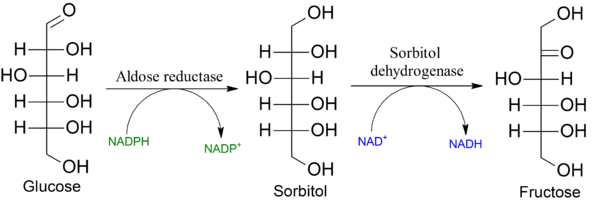
Carbohydrates with high glycaemic index are proposed to promote the development of obesity, insulin resistance and fatty liver, but the mechanism by which this occurs remains unknown. High serum glucose concentrations are known to induce the polyol pathway and increase fructose generation in the liver. Here we show that this hepatic, endogenously produced fructose causes systemic metabolic changes. We demonstrate that mice unable to metabolize fructose are protected from an increase in energy intake and body weight, visceral obesity, fatty liver, elevated insulin levels and hyperleptinaemia after exposure to 10% glucose for 14 weeks. In normal mice, glucose consumption is accompanied by aldose reductase and polyol pathway activation in steatotic areas. In this regard, we show that aldose reductase-deficient mice are protected against glucose-induced fatty liver. We conclude that endogenous fructose generation and metabolism in the liver represents an important mechanism by which glucose promotes the development of metabolic syndrome.

The Polyol Pathway


To summarise - when extra glucose is consumed by mice, some of this is converted to sorbitol then to fructose. This is associated with elevation of AST and ALT. Mice that cannot convert convert glucose to sorbitol are protected from rises in fructose and liver enzymes. Therefore fructose is so bad for you that it even accounts for the metabolic harm of high glucose intakes.
"
These studies show that glucose-mediated obesity, visceral fat accumulation, hyperinsulinaemia, hyperleptinaemia and fatty liver are all dependent in part on the conversion of glucose to fructose in the liver with the metabolism of fructose by KHK. In other words, the mechanism by which glucose induces its metabolic effects is largely dependent on fructose
metabolites resulting 
from the fructose generated from glucose by the polyol pathway".


But, one must insist - this is dietary glucose doing this, not dietary fructose. This is not evidence in support of the idea that fructose is the bad carb, and glucose is the good carb; it looks more like evidence that, in normal metabolisms, sugars are somewhat interchangeable.
If you consume an excess of glucose, hepatic metabolism will have to convert some of that excess to fatty acids. Palmitic acid will be synthesised, and some palmitic acid will be elongated to oleic acid (because you can't make a triglyceride with 3 SFAs - if there's no dietary MUFA, DNL oleate makes the process of esterification possible). But you'll also need glycerol, and fructose is said to be a better glycerol substrate than glucose (for some reason I don't understand, being hopeless at maths). Note the pyruvate as a source of fatty acids in the schema below - this could just as easily have come from glucose. Indeed, the acetyl-CoA to make fatty acids could have come from fatty acids. (This is what happens with linoleic acid in fatty liver disease. Indeed, this kind of futile cycling of lipid carbons seems to be a feature of NAFLD.)

Fructose-triglyceride.jpg


In the context of hyperglycemia in the absence of dietary fructose, (
one might speculate that) 
the generation of a little fructose makes sense, just as the elongation of palmitate to oleate makes sense, as a means of packing away energy from glucose into stable triglycerides (which can be exported from hepatocytes with VLDL, or stored more-or-less safely for a while) more easily than would otherwise be the case. Perhaps.


We fed the mice 10% glucose water and off they went.


The knock-out mice that couldn't change glucose into fructose consumed less glucose. The authors pulled out some KO mice (n=4) that ate as much as 4 non-KO mice, to show that the KO mice were healthier - fully protected against the metabolic harm of fructose/glucose - but this kind of post-hoc tactic is a bit suspect. Interestingly the KO mice had higher serum beta-hydroxybutyrate (ketone body) levels. I guess that energy had to go somewhere (is this at all relevant to the claims that potatoes elevate ketones?).


There are humans who cannot metabolise fructose or sorbitol.

"
Affected individuals are asymptomatic and healthy, provided they do not ingest foods containing fructose or any of its common precursors, sucrose and sorbitolMost adult patients do not have any dental caries
".

Bill Lagakos says that his nutrition tutors spent all of five minutes on the polyol pathway; R.D. Feinman, who should know, states "I never saw the point or function of the polyol pathway".
Wild speculation aside, that's fair comment. The polyol pathway is useful for blinding diabetics, but it doesn't seem particularly essential for life. At most it might once have provided a slight buffer against hyperglycemia, but today we have far too many copies of the amylase gene and the polyol shock absorber, if that is what it was, is easily broken, making things worse. Is that any explanation? Some mysteries, it seems, are going to remain mysteries.


Fructose is a factor in fibrosis of chronic Hepatitis C: but it's not a biggie.

Industrial, not fruit fructose intake is associated with the severity of liver fibrosis in genotype 1 chronic hepatitis C patients.
Unhealthy food intake, specifically fructose, has been associated with metabolic alterations and with the severity of liver fibrosis in patients with non-alcoholic fatty liver disease. In a cohort of patients with genotype 1 chronic hepatitis C (G1 CHC), we tested the association of fructose intake with the severity of liver histology.

METHODS:

Anthropometric and metabolic factors, including waist circumference (WC), waist-to-hip ratio (WHR), dorso-cervical lipohypertrophy and HOMA were assessed in 147 consecutive biopsy-proven G1 CHC patients. Food intake, namely industrial and fruit fructose, was investigated by a three-day structured interview and a computed database. All biopsies were scored by an experienced pathologist for staging and grading (Scheuer classification), and graded for steatosis, which was considered moderate-severe if ⩾20%. Features of non-alcoholic steatohepatitis (NASH) in CHC were also assessed (Bedossa classification).

RESULTS:

Mean daily intake of total, industrial and fruit fructose was 18.0±8.7g, 6.0±4.7g, and 11.9±7.2g, respectively. Intake of industrial, not fruit fructose, was independently associated with higher WHR (p=0.02) and hypercaloric diet (p=0.001). CHC patients with severe liver fibrosis (⩾F3) reported a significantly higher intake of total (20.8±10.2 vs. 17.2±8.1g/day; p=0.04) and industrial fructose (7.8±6.0 vs. 5.5±4.2; p=0.01), not fruit fructose (12.9±8.0 vs. 11.6±7.0; p=0.34). Multivariate logistic regression analysis showed that older age (OR 1.048, 95% CI 1.004-1.094, p=0.03), severe necroinflammatory activity (OR 3.325, 95% CI 1.347-8.209, p=0.009), moderate-severe steatosis (OR 2.421, 95% CI 1.017-6.415, p=0.04), and industrial fructose intake (OR 1.147, 95% CI 1.047-1.257, p=0.003) were independently linked to severe fibrosis. No association was found between fructose intake and liver necroinflammatory activity, steatosis, and the features of NASH.

CONCLUSIONS:

The daily intake of industrial, not fruit fructose is a risk factor for metabolic alterations and the severity of liver fibrosis in patients with G1 CHC.


Excuse me, but OR 1.147 is not a huge correlation. It's as tiny as a red-meat-and-disease-of-your-choice correlation in a study run by vegans. What if they had looked at total carbohydrate? Or included fruit juice and high-GI glucose sources like bread and pasta? Do you think the OR would have been higher then? It was about OR 2.9 for carbohydrate and fibrosis in the last Italian diet study I read.
Yes, my header does say limit fructose. And fructose might have special powers to harm, it's just hard to distinguish how strong those are when the trend is to put all the blame on them. The usual sources of fructose - sugar, HFCS, fruit juice - are mainly dumping carbohydrate directly into circulation, like that 10% glucose water the mice drank. They are high GI carbs. Or they would be if F was also G. But F is also G, and G is also F. 
If we can learn anything from the polyol pathway, it is, that carbs will be carbs.


P.S. remember from the NASH posts how taurine is good for removing excess cholesterol from the liver? And generally hepatoprotective?

Sorbitol accumulation depletes taurine:

Prevention of sorbitol accumulation with the aldose reductase inhibitor sorbinil increased nerve taurine levels by 22% (p < 0.05) when compared with untreated diabetic animals. Thus, we have demonstrated an interdependence of organic osmolytes within the nerve. Abnormal accumulation of one osmolyte results in reciprocal depletion of others. Diabetic neuropathy may be an example of maladaptive osmoregulation, nerve damage and instability being aggravated by taurine depletion.